Synthesis and characterization of NM-PB Nanozymes
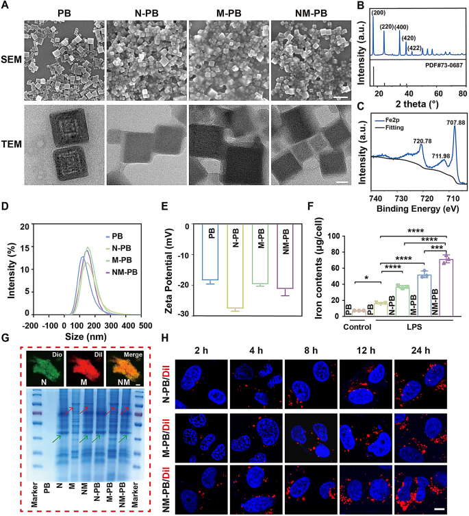
The synthesis of PB nanoparticles is described intimately in technique. The morphology and measurement of PB nanozyme could be clearly noticed via the utilization of SEM and TEM methods (Fig. 1A). The outcomes point out that the PB nanozyme reveals a well-defined cubic crystal construction. The X-ray diffraction (XRD) patterns reveal distinct diffraction planes of PB at 17.5° (200), 24.8° (220), 35.4° (400), and 39.7° (420) as proven in Fig. 1B. The chemical state of PB was characterised utilizing X-ray photoelectron spectroscopy (XPS). As depicted in Fig. 1C, the peaks noticed at Fe2P3/2 (711.98 eV) and Fe2P1/2 (720.78 eV) correspond to the presence of FeIII in Fe3[Fe2(CN)6]4, whereas the height at 707.88 eV signifies the existence of Fe2P3/2 in [Fe2 (CN) 6]4−.
To visualise the membrane fusion, a 1:1 protein weight ratio of Dio-labeled Neutrophil membranes (N) and Dil-labeled macrophage membranes (M) was ready and sonicated at 37 °C for 10 min. Utilizing the BCA technique and inductively coupled plasma optical emission spectrometry (ICP-OES), the ratio of the cell membrane to PB was calculated. The outcomes confirmed that the neutrophil-macrophage hybrid membrane accounts for 31.5% of the NM-PB nanozyme, whereas Prussian blue accounts for 68.5%. CLSM revealed a major co-localization of the fluorescent alerts originating from the NM hybrid membrane (Fig. 1G). Then, the identical focus of PB nanozymes (200 µg/mL) was combined with an equal quantity of cell membranes, and the combined answer was sonicated in an ice tub for 10 min to kind N-PB, M-PB, NM-PB nanozymes by the ultrasonic technique. Moreover, Coomassie sensible blue staining evaluation revealed that the protein bands noticed on NM-PB have been corresponding to these of remoted N and M fractions, as indicated by the inexperienced and purple arrows, thereby indicating the presence of membrane proteins (Fig. 1G). The presence of skinny movie coatings on the surfaces of NM-PB, N-PB, and M-PB nanozymes was confirmed via direct observations through TEM and SEM. As depicted in Fig. 1D-E, Dynamic gentle scattering (DLS) evaluation confirmed that the common particle measurement of PB is 108.3 nm. Nonetheless, after modification with cell membranes, the sizes of the nanozymes elevated considerably: N-PB to 156.5 nm, M-PB to 154.1 nm, and NM-PB to 159.0 nm. The Zeta potential measurements revealed that NM-PB (-19.0 mV), N-PB (-28.4 mV), and M-PB (-23.7 mV) nanozymes exhibit a considerably greater adverse cost in comparison with the unmodified PB (-17.1 mV). The presence of a powerful adverse zeta potential on the floor of cell membranes is liable for this phenomenon, indicating the profitable modification of PB nanozymes with mobile membranes.
Characterization of NM-PB nanozymes and focusing on capability of NM-PB nanozymes in vitro. (A) SEM photographs (scale bar = 200 nm) and TEM photographs (scale bar = 20 nm) of PB, N-PB, M-PB, NM-PB nanozymes. (B) XRD sample of PB nanozymes. (C) XPS spectra of PB nanozymes within the Fe 2p area. D–E) DLS and Zeta potential evaluation of PB, N-PB, M-PB, and NM-PB PB nanozymes. F) The concentrations of iron measured by ICP-OES in numerous teams (n = 3). G) CLSM photographs of the neutrophil-macrophage hybrid membrane (scale bar = 10 μm); SDS-PAGE protein evaluation of PB nanozymes, N, M, NM, N-PB nanozymes, M-PB nanozymes, and NM-PB nanozymes by Coomassie blue staining. H) Intracellular uptake of N-PB, M-PB, and NM-PB nanozymes (the blue signifies nucleus stained with DAPI, the purple signifies N-PB, M-PB, and NM-PB nanozymes stained with Dil) (scale bar = 20 μm). *p < 0.05, ***p < 0.001, ****p < 0.0001. one-way ANOVA, Tukey’s a number of comparability check
In vitro biocompatibility and focusing on functionality of NM-PB nanozymes
The non-toxicity of nanomedicine is an important prerequisite for its profitable medical translation. To evaluate the biocompatibility of NM-PB nanozymes and examine its potential anti-inflammatory results in vitro, we carried out cytotoxicity assays using FHC cells and RAW 264.7 immune cells. After exposing FHC cells and RAW 264.7 cells to various concentrations of NM-PB nanozymes for twenty-four h, even at excessive concentrations of PB nanozymes (200 µg/mL), cell viability remained above 90% (Fig. S1A-B). These outcomes verify that NM-PB nanozymes exhibit wonderful biocompatibility, positioning them as viable candidates for drug supply functions in nanotherapeutics.
To additional examine the focusing on functionality of NM-PB nanozymes, we established a colitis cell mannequin [32]. The focusing on efficacy of NM-PB nanozymes on FHC cells was subsequently evaluated. The cell membrane was labeled with Dil fluorescent dye for CLSM visualization. Cells have been incubated with N-PB/Dil, M-PB/Dil, and NM-PB/Dil nanozymes (at an PB focus of 200 µg/mL) for numerous durations (2 h, 4 h, 8 h, 12 h, and 24 h). CLSM imaging revealed considerably larger intracellular accumulation of nanoparticles within the NM-PB group in comparison with the N-PB and M-PB teams (Fig. 1H, Fig. S2A). The iron content material in cells was decided utilizing ICP-OES to additional validate this discovering. In comparison with the PB group, N-PB group, and M-PB group, the NM-PB group exhibited the very best iron content material in colitis cell fashions. This additional signifies that NM-PB nanozymes enhanced the uptake of PB nanozymes (Fig. 1F). It’s evident that nanozymes modified with two forms of cell membranes possess twin focusing on skills, which consequently enhances their enrichment and uptake capability on the lesion website.
Evaluation of the ROS scavenging capability of NM-PB nanozymes in vitro
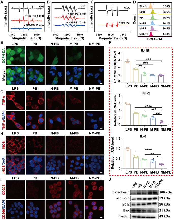
The Electron Spin Resonance (ESR) method is also used for the exact detection of free radicals because of its distinctive sensitivity [34]. Consequently, the measurement of inflammation-related species resembling •OH, •OOH, and H2O2 following NM-PB nanozymes administration was additional performed utilizing ESR. The ESR outcomes demonstrated a major discount within the attribute peak intensities of BMPO /•OH, BMPO/•OOH and TEMPO/ H2O2 with the applying of NM-PB nanozymes (Fig. 2A-C).
The extreme manufacturing of ROS in cells, induced by a state of complete oxidative stress, triggers a cascade of protein kinases in inflammatory tissues, thereby selling oxidative injury [10]. The potent antioxidant properties of NM-PB nanozymes prompted us to research the potential of NM-PB nanozymes in restoring cell survival beneath circumstances of excessive oxidative stress, in addition to assessing the in vitro capability of NM-PB nanozymes to remove ROS. The LPS group is a colitis cell mannequin established by LPS-induced FHC cells, adopted by remedy with PB, N-PB, M-PB, and NM-PB nanozymes at equal doses, respectively. Subsequently, DCFH-DA staining was employed to evaluate ROS ranges. As depicted in Fig. 2E and Fig. S2B, a major improve in inexperienced fluorescence sign was noticed in FHC cells inside the LPS group. Notably, remedy with PB, N-PB, M-PB, and NM-PB nanozymes resulted in lowered ROS ranges in comparison with the LPS group, with NM-PB nanozymes exhibiting essentially the most pronounced ROS scavenging functionality amongst all examined formulations. We additional performed quantitative evaluation of intracellular ranges of ROS utilizing move cytometry, which is in keeping with the earlier outcomes (Fig. 2D). These findings point out that PB nanozymes, possessing antioxidant enzyme exercise, successfully eliminates ROS and protects cells from oxidative stress. Importantly, the antioxidant impact was significantly pronounced within the NM-PB group, suggesting that the twin focusing on capacity of neutrophil-macrophage hybrid membrane enhances mobile uptake of PB nanozymes and thereby augments its antioxidant efficacy.
The colonic epithelial cells in UC lesions are subjected to wreck brought on by ROS, resulting in the next launch of a major quantity of damage-associated molecular patterns (DAMPs) into the intercellular area. These DAMPs stimulate the differentiation of macrophages right into a pro-inflammatory phenotype generally known as M1 macrophages, which secrete numerous pro-inflammatory cytokines. These cytokines can additional improve intracellular ROS manufacturing, thereby establishing a detrimental cycle of irritation and oxidative stress [35, 36].
The anti-inflammatory efficacy of NM-PB was confirmed by measuring cytokines related to irritation utilizing RT-qPCR. As depicted in Fig. 2F, the mRNA expression ranges of IL-1β, TNF-α, and IL-6 exhibited a major lower following remedy with PB, N-PB, M-PB, and NM-PB nanozymes when in comparison with the LPS group. Notably, NM-PB nanozymes demonstrated a very pronounced anti-inflammatory impact. Moreover, mobile immunofluorescence evaluation revealed constant findings relating to the adjustments in TNF-α inflammatory issue inside the colitis cell mannequin. Remarkably lowered expression of TNF-α was noticed after NM-PB nanozymes remedy (Fig. 2G, Fig. S2C). These outcomes collectively point out that NM-PB nanozymes possesses potent anti-inflammatory capabilities.
The apoptosis of intestinal epithelial cells in UC could be attributed to the secondary injury inflicted on intestinal cells by ROS [37]. The anti-apoptotic functionality of NM-PB nanozymes was confirmed by evaluating apoptosis-related biomarkers through Western Blot (WB). The expression of the pro-apoptotic protein Bax was considerably suppressed within the N-PB, M-PB, and NM-PB teams in comparison with the LPS group, as proven in Fig. 2J and Fig. S2D. Conversely, an upregulation within the expression of the anti-apoptotic protein Bcl2 was noticed (Fig. 2J, Fig. S2E). Notably, amongst these teams, the NM-PB group exhibited superior anti-apoptotic functionality. Due to this fact, it may be inferred that NM-PB nanozymes successfully mitigates ROS-induced cell apoptosis.
Based mostly on the shut correlation between intestinal barrier dysfunction and the pathogenesis of UC [38]. The tight junction and adherens junction proteins, together with ZO-1, occludin and E-cadherin [39]. We hypothesize NM-PB nanozymes can restore the impaired intestinal barrier operate within the LPS-induced mobile colitis mannequin. Subsequently, numerous remedies together with PB, N-PB, M-PB, and NM-PB nanozymes have been administered to the colitis mobile mannequin. The WB evaluation outcomes confirmed that, in contrast with the PB, N-PB, and M-PB teams, the upregulation of E-cadherin and Occludin expression was most pronounced within the NM-PB group (Fig. 2J, Fig. S2F-G).
Activated inflammatory macrophages play a pivotal position within the pathophysiology of intestinal colitis [40]. LPS-stimulated RAW264.7 macrophages endure activation characterised by the upregulation of inducible nitric oxide synthase (iNOS), a trademark of M1 macrophage polarization, which ends up in the manufacturing of great portions of reactive nitrogen species (RNS), together with nitric oxide (NO). These RNS can disrupt mitochondrial oxidative phosphorylation, leading to an as much as tenfold improve within the launch of ROS [41].
To additional elucidate the potential of NM-PB nanozymes in selling the polarization of M1 macrophages in direction of an M2 phenotype, we performed mobile immunofluorescence assays. Our findings revealed that PB, N-PB, M-PB, and NM-PB nanozymes exhibited a exceptional capacity to attenuate LPS-induced activation of M1 macrophages and downregulate iNOS expression when in comparison with the LPS group. Moreover, these remedies upregulated the expression of CD206, a floor protein marker related to M2 macrophages. Notably, NM-PB nanozymes demonstrated essentially the most important impact in inducing this transition (Fig. 2H-I, Fig. S2H-I).
These findings point out that NM-PB nanozymes possesses the aptitude to disrupt the detrimental suggestions loop established by ROS and extreme immune response within the colitis cell mannequin, thereby mitigating apoptosis of intestinal epithelial cells and restoring intestinal mucosal barrier operate. Moreover, it will possibly facilitate the conversion of M1 macrophages into M2 phenotype. This impact could also be attributed to the dual-targeting functionality of neutrophil-macrophage hybrid membrane, which reinforces intracellular accumulation of PB nanozymes and additional improves their useful efficacy.
ROS scavenging exercise of NM-PB nanozymes and induction of macrophage reprogramming in vitro. A–C) The scavenging impact of NM-PB nanozymes on •OH, •OOH, and H2O2. D–E) Intracellular ROS scavenging capability of NM-PB nanoenzymes as detected by move cytometry and CLSM (scale bar = 10 μm). F) Relative mRNA ranges of pro-inflammatory cytokines (IL-1β, IL-6, and TNF-α) in FHC cells handled with totally different remedies detected by RT-qPCR (n = 3). G) Immunofluorescence staining of TNF-α in FHC cells after totally different remedies (scale bar = 10 μm). H–I) Immunofluorescence staining of iNOS and CD206 in RAW264.7 cells after totally different remedies (scale bar = 10 μm). J) Western blot evaluation of the expression of E-cadherin, Occludin, Bcl2, and Bax proteins in FHC cells after totally different remedies. *p < 0.05, **p < 0.01, ***p < 0.001, ****p < 0.0001. one-way ANOVA, Tukey’s a number of comparability check
Evaluation of the therapeutic efficacy of PB nanozymes in UC
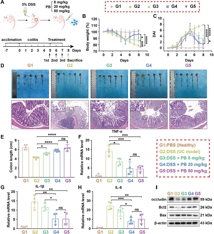
The potent ROS scavenging exercise of PB in vitro warrants additional investigation into its therapeutic efficacy in a murine mannequin of DSS-induced UC. all mice have been randomly divided into 5 teams (n = 5): G1: PBS group (wholesome mice receiving PBS), G2: DSS group (mice with UC), G3: DSS + PB 8 mg/kg group, G4: DSS + PB 20 mg/kg group, G5: DSS + PB 50 mg/kg group. Mice have been administered with 3% DSS for 5 days to induce an acute UC mannequin, adopted by intravenous tail injections of PB nanozymes on days 5, 6, and seven. The optimum therapeutic dose of PB nanozymes was decided by treating the UC mouse mannequin with totally different concentrations of PB nanozymes (8 mg/kg, 20 mg/kg, 50 mg/kg) (Fig. 3A).
To evaluate the protecting results of PB nanozymes on UC lesions, we measured the physique weight, colon size, and illness exercise index (DAI) of the mice, using the strategy described in earlier literature [42], and performed histological examinations of the distal colon. The outcomes depicted in Fig. 3B-E reveal that mice handled with various doses of PB nanozymes exhibited important enhancements in physique weight, intestinal tube size, and DAI in comparison with G2 group. The HE staining outcomes revealed that the crypts of the intestinal tissue within the G2 group exhibited full distortion, extreme lack of goblet cells, and evident infiltration of inflammatory cells (Fig. S2J). In distinction, these pathological adjustments have been mitigated within the G3, G4, and G5 teams. Apparently, the therapeutic results of G4 and G5 teams have been comparable, with each considerably mitigating inflammatory injury in mice.
Subsequently we investigated the impact of PB nanozymes on the expression of inflammatory cytokines in mice UC mannequin. RT-qPCR evaluation revealed that in comparison with the G2 group, the mRNA expression ranges of TNF-α, IL-1β, and IL-6 have been mitigated in G3, G4, and G5 teams. (Fig. 3F-H). Once more, the results within the G4 and G5 teams have been comparable, additional supporting the anti-inflammatory efficacy of PB nanozymes in assuaging DSS-induced UC.
To discover whether or not PB can restore epithelial barrier in mice with UC, various concentrations of PB (8 mg/kg, 20 mg/kg, 50 mg/kg) have been administered to the colitis mouse mannequin. The WB outcomes demonstrated the expression ranges of Occludin was considerably upregulated within the G3, G4, and G5 teams, as in comparison with the G2 group (Fig. 3I, Fig. S2Okay). Moreover, the induction of ROS has been related to elevated expression of the pro-apoptotic protein Bax, which promotes apoptosis of intestinal epithelial cells [37]. Following remedy with PB nanozymes, we noticed a major downregulation of Bax and an upregulation of the anti-apoptotic protein Bcl-2 (Fig. 3I, Fig. S2L-M). Notably, each the G4 and G5 teams exhibited comparable results in mitigating apoptosis of intestinal epithelial cells.
These findings point out that PB nanozymes possess the capability for anti-inflammatory, anti-apoptotic results, and restoring the performance of the intestinal epithelial barrier. Given the comparable efficacy noticed between the G4 and G5 teams, we chosen a dosage of 20 mg/kg for subsequent in vivo experiments.
Willpower of the optimum dosage of PB nanozymes for the remedy of UC mannequin in mice. A) Schematic illustration of the experimental protocol employed for UC remedy utilizing PB nanozymes. B) Modifications in physique weight following numerous remedies. C) Illness Exercise Index (DAI) adjustments after totally different remedies. D) Consultant photographs illustrating the macroscopic look and H&E staining of colon tissue after totally different remedies (scale bar = 200 μm). E) Colon size (n = 5). F–H) The expression of mRNAs for TNF-α, IL-1β, and IL-6 was decided by RT-qPCR after totally different remedies (n = 4). I) The expression of proteins for Occludin, Bcl2, and Bax was analyzed by Western blot after totally different remedies. ns, not important. *p < 0.05, **p < 0.01, ***p < 0.001, ****p < 0.0001. one-way ANOVA, Tukey’s a number of comparability check
Biocompatibility of NM-PB nanozymes
Previous to in vivo software, we assessed the biosafety of NM-PB nanozymes via evaluation of hematological markers and histopathology examination of significant organs. The administration of NM-PB nanozymes didn’t result in any important abnormalities in liver operate, renal operate, or blood routine parameters, as demonstrated in Fig. S3A-F. Moreover, histological evaluation utilizing hematoxylin and eosin (H&E) staining demonstrated no important pathological alterations in main organs, together with the guts, liver, spleen, lungs, and kidneys, following remedy with NM-PB nanozymes (Fig. S3G). The findings validate the whole absorption and metabolism of NM-PB nanozymes after intravenous injection, demonstrating its favorable biosafety profile. This helps their potential for in vivo functions and gives a possible pathway for translating remedy from animal experiments to medical functions.
In vivo focusing on functionality and therapeutic efficacy of NM-PB nanozymes
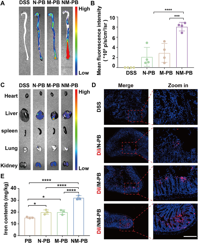
The correct supply of nanozymes to UC lesion website is essential for guaranteeing therapeutic efficacy. Whereas we have now beforehand demonstrated the in vitro focusing on functionality of NM-PB nanozymes, the present examine focuses on evaluating their localization at UC lesions in a murine mannequin. To facilitate visualization, the cell membranes of the nanozymes have been labeled with Dir fluorescent dye. Following the intravenous administration of equimolar quantities of N-PB, M-PB, and NM-PB nanozymes into the mouse UC mannequin, the colons have been harvested at 24 h for fluorescence imaging to visualise their distribution. As depicted in Fig. 4A-B, the fluorescence sign was noticed to be minimal within the N-PB and M-PB teams, respectively. whereas a strong fluorescence sign was detected within the NM-PB group, indicating the numerous focusing on functionality of the neutrophil-macrophage hybrid membrane. Moreover, the biodistribution of the injected nanozymes in main organs was additionally evaluated. Biodistribution evaluation revealed fluorescent alerts within the liver and kidneys 24 h post-intravenous administration (Fig. 4C, Fig. S4A), suggesting these organs play a task within the clearance of nanozymes throughout systemic circulation.
The in vivo focusing on functionality of NM-PB nanozymes to UC lesions was investigated utilizing CLSM subsequently. The cell membrane was labeled with Dil fluorescent dye for CLSM visualization. After a 24 h interval following intravenous administration, frozen sections of diseased intestinal tissue have been examined utilizing CLSM. A small amount of purple fluorescence sign was detected on the website of colon lesions in each the N-PB and M-PB teams. In distinction, a major enhancement of alerts was noticed within the NM-PB group (Fig. 4D, Fig. S4B), corroborating the ex vivo fluorescence imaging outcomes.
The enrichment of nanozymes in intestinal lesions was additional quantified by analyzing the iron content material utilizing ICP-OES. In comparison with the PB, N-PB, and M-PB teams, the NM-PB group exhibited a considerably greater focus of iron within the diseased intestinal tissues (Fig. 4E), additional confirming the superior focusing on capabilities of NM-PB nanozymes in direction of UC lesions. Collectively, these outcomes reveal that neutrophil-macrophage hybrid membrane reveals wonderful focusing on functionality to UC lesions.
In vivo focusing on capability of NM-PB nanozymes. A) Ex vivo fluorescence imaging of colon tissues from every group. B) Quantitative evaluation of imply fluorescence depth in colon tissues (n = 4). C) Ex vivo fluorescence imaging of predominant organs. D) CLSM photographs of autofluorescence in colon tissue sections (The blue fluorescence signifies the presence of nuclei, whereas the purple fluorescence corresponds to Dil-labeled N-PB, M-PB, and NM-PB nanozymes; scale bar = 500 μm). E) Quantification of iron concentrations measured by ICP-OES following numerous remedies (n = 3). *p < 0.05, ***p < 0.001, ****p < 0.0001. one-way ANOVA, Tukey’s a number of comparability check
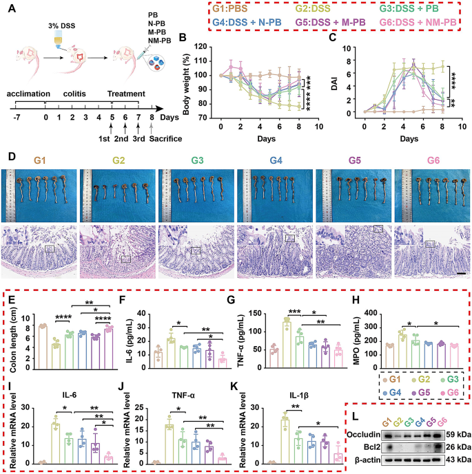
To additional evaluate the therapeutic efficacy of PB, N-PB, M-PB, and NM-PB nanozymes in UC, all mice have been randomly divided into six teams: G1: PBS group (wholesome mice receiving PBS), G2: DSS group (mice with UC), G3: DSS + PB group, G4: DSS + N-PB group, G5: DSS + M-PB group, and G6: DSS + NM-PB group. The therapeutic efficacy was evaluated by assessing adjustments in physique weight, DAI, colon size, expression ranges of proinflammatory cytokines, and histological evaluation of colon sections among the many teams. In comparison with the G2 group, the G3, G4, G5, and G6 teams all demonstrated considerably longer colon lengths, greater physique weights, and decrease DAI scores (Fig. 5A-E), with essentially the most pronounced enchancment noticed within the G6 group. The histomorphological evaluation of the colon revealed that G2 Group displayed extreme crypt destruction, in depth infiltration of immune cells, and important injury to colonic epithelial cells. In stark distinction, G6 Group displayed practically regular histological microstructures (Fig. 5D, Fig. S4C). The outcomes reveal that the therapeutic modality within the G6 Group considerably ameliorated each the signs and histomorphological options of the colitis mice mannequin.
The outcomes from RT-qPCR and ELISA assays reveal that, in comparison with the G2 Group, the G3, G4, G5, and G6 Teams considerably inhibited the expression of inflammatory cytokines IL-6, TNFα, IL-1β, and myeloperoxidase (MPO). Notably, the G6 Group exhibited a superior impact in inhibiting the pro-inflammatory cytokines (Fig. 5F-Okay). These outcomes could be attributed to the dual-targeting motion of NM-PB, which reinforces the buildup of PB at UC lesion websites, thereby considerably enhancing its anti-inflammatory impact.
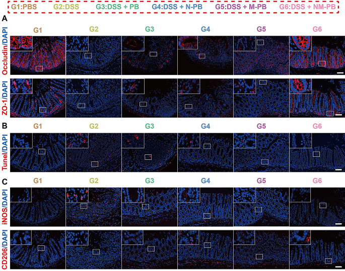
The WB and tissue immunofluorescence analyses revealed that the expression of ZO-1 and Occludin was considerably upregulated within the inflammatory lesions of the G6 group in comparison with the G3, G4, and G5 teams (Figs. 5L and 6A, Fig. S4D, Fig. S4F-G). These outcomes present compelling proof that NM-PB nanozyme remedy enhances the expression of tight junction proteins, facilitating the meeting of tight junction complexes and successfully restoring intestinal barrier operate.
Therapeutic efficacy of NM-PB nanozymes in mice UC mannequin. A) Schematic illustration of the experimental protocol employed for the remedy of UC utilizing PB, N-PB, M-PB, NM-PB nanozymes. B) Physique weight adjustments after totally different remedies. C) DAI adjustments after totally different remedies. D) Consultant photographs of macroscopic look and H&E staining of colon tissues (scale bar = 200 μm). E) Colon size (n = 6). F–H) The degrees of IL-6, TNF-α, and MPO as decided by ELISA within the colon tissues after totally different remedies (n = 5). I–Okay) The expression of mRNAs for IL-6, TNF-α, and IL-1β as decided by RT-qPCR within the colon tissues after totally different remedies. L) The expression of proteins for Occludin and Bcl2 as analyzed by Western blot within the colon tissues after totally different remedies. *p < 0.05, **p < 0.01, ***p < 0.001, ****p < 0.0001. one-way ANOVA, Tukey’s a number of comparability check
TUNEL staining moreover demonstrated the incidence of mobile apoptosis in colon tissue. Notably, the purple fluorescence depth related to TUNEL staining within the G6 group confirmed a major lower in comparison with the G3, G4, and G5 teams (Fig. 6B, Fig. S4H). Correspondingly, the expression stage of Bcl2 protein within the G6 group markedly elevated after remedy, indicating that NM-PB nanozymes can successfully alleviate DSS-induced apoptosis of intestinal epithelial cells (Fig. 5L, Fig. S4E). Moreover, the expression of iNOS was considerably downregulated in group G6 in comparison with teams G3, G4, and G5. In distinction, the expression of CD206 was notably upregulated within the G6 group. These findings point out that NM-PB nanozymes successfully facilitates the polarization of macrophages from the M1-phenotype to the M2-phenotype, thereby offering safety in opposition to oxidative stress for intestinal epithelial cells (Fig. 6C, Fig. S4I-J). The hybrid membrane-modified supply system makes use of the inflammatory chemotactic properties of neutrophil and macrophage membranes, selling the buildup of PB nanozymes in UC lesions. This focused supply method permits the efficient exploitation of the varied functionalities of PB nanozymes, which embrace anti-inflammatory results, anti-apoptotic actions, restoration of the intestinal mucosal barrier, and promotion of M1 macrophage polarization in direction of the M2 phenotype.
NM-PB nanozymes restore mucosal barrier operate and promote macrophage reprogramming in mice with UC. A) Immunofluorescence staining of ZO-1 and Occludin in colon tissues after totally different remedies (scale bar = 200 μm). B) Consultant photographs of TUNEL staining in colon tissues after totally different remedies (scale bar = 200 μm). C) Immunofluorescence staining of iNOS and CD206 expression in colon tissues after totally different remedies (scale bar = 200 μm)
Given the restrictions of conventional medicine for treating UC, we in contrast the biosafety and therapeutic efficacy of NM-PB nanozyme and 5-ASA in a mouse mannequin of UC. all mice have been randomly divided into three teams: (1) DSS (mice with DSS-induced colitis with out remedy, n = 6), (2) 5-ASA (mice with DSS-induced colitis handled with 5-ASA, n = 5), (3) NM-PB (mice with DSS-induced colitis handled with NM-PB nanozymes, n = 5). The outcomes proven in Fig. S5A-G reveal that NM-PB nanozyme and 5-ASA exhibit good biosafety in UC mouse fashions. In comparison with the DSS group, each the 5-ASA and NM-PB teams demonstrated longer colon lengths, greater physique weights, and decrease DAI scores. Moreover, mice in each the 5-ASA and NM-PB teams displayed practically regular histological microstructures (Fig. S6A-F), with essentially the most pronounced enchancment noticed within the NM-PB group. In comparison with colitis mice handled with 5-ASA, NM-PB nanozyme is simpler in inhibiting inflammatory responses (Fig. S6G-I), lowering apoptosis of intestinal epithelial cells (Fig. S6J, Fig. S7C), restoring the integrity of the intestinal mucosal barrier (Fig. S6J, Fig. S7A-B), and selling the polarization of macrophages from an M1-phenotype in direction of an M2-phenotype (Fig. S7D-E). Due to this fact, in comparison with colitis mice handled with 5-ASA alone, NM-PB nanozyme is anticipated to supply higher therapeutic advantages for assuaging UC.
Transcriptomic evaluation of the therapeutic mechanism of NM-PB nanozymes in vivo
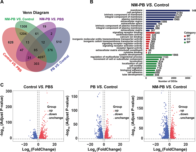
To elucidate the therapeutic mechanism of NM-PB nanozymes in vivo and uncover the molecular mechanisms in addition to signaling pathways that contribute to its effectiveness in treating UC, we performed RNA sequencing on colonic tissues from 4 experimental teams: PBS group, DSS group, DSS + PB group, and DSS + NM-PB group. The appliance of Principal Element Evaluation (PCA) unveiled distinct transcriptomic profiles among the many 4 teams (Fig. S8A). Moreover, distinctive transcriptomic traits have been evident from the Venn diagrams and heatmaps (Fig. 7A, Fig. S8B).
The volcano plot and the bar chart of complete DEGs point out that, in comparison with the PBS group, the DSS group exhibited 7,008 DEGs, with 4096 genes upregulated and 2912 genes downregulated. After remedy with PB Nanozymes, a complete of 6016 DEGs (2627 upregulated genes and 3389 downregulated genes) have been recognized. Remarkably, following remedy with NM-PB nanozymes, a complete of 7756 DEGs have been noticed, comprising 2959 upregulated genes and 4797 downregulated genes (Fig. 7C, Fig. S8C). Notably, the transcriptome profile of NM-PB-treated mice with UC resembles that of the PBS group.
Subsequently, we carried out clustering and enrichment analyses on the DEGs to make clear the therapeutic mechanisms of NM-PB nanozymes. The DEGs have been initially categorized into three teams, particularly molecular operate (MF), organic course of (BP), and mobile element (CC), primarily based on the gene ontology database (GO). There have been important variations (p < 0.05) in a number of organic pathways between the DSS group and NM-PB group, together with cytokine binding, oxidoreductase exercise, sign receptor binding, and sign receptor regulator exercise (Fig. 7B).
RNA-seq evaluation of DSS-induced colitis regulated by NM-PB nanozymes. A) Venn diagram illustrating the differentially expressed genes (DEGs) recognized via complete transcriptome RNA-seq evaluation among the many PBS, DSS, PB, and NM-PB teams (| log2(fold change) | > 1, adjusted P < 0.05). B) GO useful enrichment in NM-PB group Vs. DSS group. C) Volcano plot exhibiting the DEGs within the DSS group Vs. the PBS group (left), within the PB group Vs. DSS group (center), within the NM-PB group Vs. DSS group (proper) from RNA-seq information (up-regulated genes: purple; down-regulated genes: blue)
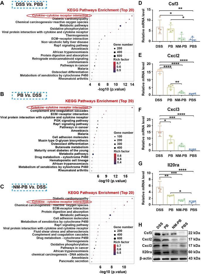
The Kyoto Encyclopedia of Genes and Genomes database (KEGG) [43] was carried out to research associated objects with the log2FC expression stage of DEGs because the analysis commonplace. Between the DSS group versus PBS group, PB group versus DSS group, and NM-PB group versus DSS group, DEGs have been all related to the cytokine-cytokine receptor interplay signaling pathway in KEGG (Fig. 8A-C).
Due to this fact, we speculate whether or not NM-PB nanozymes regulate the development of UC via the cytokine-cytokine receptor interplay pathway. Based mostly on the evaluation of DEGs information, the expression of key molecules (Cxcl3, Il19, Il20ra) related to the cytokine-cytokine receptor interplay pathway have been inhibited (Fig. S8D-F). Outcomes from RT-qPCR and WB experiments persistently confirmed that in comparison with the DSS group, the expression ranges of Cxcl2, Cxcl3, Il20ra, and Csf3 are considerably downregulated after PB and NM-PB remedy, with the NM-PB group exhibiting expression ranges near these of the PBS group (Fig. 8D-H). In abstract, NM-PB nanozymes primarily alleviate signs in a mouse mannequin of UC by inhibiting the cytokine-cytokine receptor interplay pathway. This motion disrupts the vicious cycle of irritation and reactive oxygen species (ROS), resulting in lowered apoptosis, diminished irritation, and restored intestinal barrier operate.
Therapeutic mechanisms of the NM-PB nanozymes in mice UC mannequin. A–C) KEGG pathways enrichment evaluation of DEGs within the DSS group Vs. the PBS group, PB group Vs. the DSS group, and the NM-PB group Vs. the DSS group. D–G) The expression of mRNAs for Csf3, Cxcl2, Cxcl3, and Il20ra as decided by RT-qPCR within the colon tissues after totally different remedies. H) Validation of the expression of Csf3, Cxcl2, Cxcl3, and Il20ra in colon tissues after totally different remedies, measured by RT-qPCR and Western blot. *p < 0.05, **p < 0.01, ***p < 0.001, and ****p < 0.0001. one-way ANOVA, Tukey’s a number of comparability check


