Design and synthesis of LNT-UsSeNPs
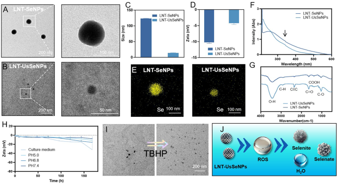
Firstly, we efficiently synthesized two varieties of Se nanoparticles: LNT-SeNPs and LNT-UsSeNPs. Uniformly dispersed LNT-SeNPs with a median measurement of ~ 100 nm had been revealed by transmission electron microscopy (TEM) photographs. Conversely, good monodispersity and a spherical form had been confirmed by LNT-UsSeNPs, which had been notably smaller, averaging 10 nm in diameter (Fig. 1A and B). Moreover, the outcomes of hydrated particle measurement and zeta potential evaluation confirmed that LNT-UsSeNPs have a median particle diameter of 13.00 ± 1.00 nm, significantly smaller than LNT-SeNPs, which have a median measurement of 123.25 ± 1.06 nm. The zeta potential of LNT-SeNPs was − 10.21 ± 0.35 mV, whereas LNT-UsSeNPs exhibited a zeta potential of -4.26 ± 0.39 mV (Fig. 1C and D). Subsequent, we assessed the steadiness of the nanoparticles by monitoring adjustments of their zeta potential in numerous options. The zeta potential of LNT-SeNPs remained secure between − 3 and − 5 mV for as much as 96 h, after which a rise in unfavorable potential indicated improve in stability. This sample was notably evident in a saline resolution (pH 5.0). In the meantime, the zeta potential of LNT-UsSeNPs remained secure between − 5 and − 10 mV for as much as 96 h, though a rise in unfavorable values was noticed at 96 h in pH 5.0, 6.8, and Dulbecco’s modified Eagle medium (DMEM). Nonetheless, in a pH 7.4 buffer saline resolution, the zeta potential of LNT-UsSeNPs remained secure between − 6 and − 8 mV, indicating enhanced stability beneath physiological situations and highlighting potential biomedical functions (Fig. 1H, S1, Supporting Info). Furthermore, EDS evaluation indicated that the principle structural element of each LNT-SeNPs and LNT-UsSeNPs was Se (Fig. 1E). The chemical buildings of LNT-SeNPs and LNT-UsSeNPs had been characterised utilizing Fourier-transform infrared spectroscopy (FT-IR) and ultraviolet-visible (UV-Vis) absorption spectroscopy. UV-Vis absorption spectroscopy confirmed that each LNT-SeNPs and LNT-UsSeNPs had excessive absorption peaks at 250 nm and 350 nm, respectively. Moreover, the stretching vibrations had been noticed at 3420 cm⁻¹ for O-H, 2920 cm⁻¹ for C-H, and 1380 cm⁻¹ for carboxyl teams, which appeared within the spectra of each LNT-SeNPs and LNT-UsSeNPs, confirmed that LNT was efficiently modified on the floor of UsSeNPs and SeNPs (Fig. 1F and G). Furthermore, LNT-UsSeNPs had been degraded after a 3 h incubation in a 1% tert-butyl hydroperoxide (TBHP) atmosphere, suggesting that LNT-UsSeNPs can react with TBHP. These outcomes indicated that LNT-UsSeNPs, owing to their smaller particle measurement and nanoscale dimensions, exhibited superior antioxidant properties and will rapidly take away TBHP (Fig. 1I).
Evaluation of the ROS scavenging capability of LNT-UsSeNPs
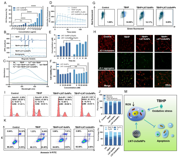
Selenium-based medicine and supplies are generally acknowledged as efficient antioxidants that help in reversing SCI attributable to dangerous ROS. Thus, an ABTS scavenging assay was used to evaluate the extracellular ROS scavenging capability of LNT-SeNPs and LNT-UsSeNPs. The outcomes indicated that LNT-UsSeNPs exhibited the next ABTS scavenging fee than LNT-SeNPs, with a transparent concentration-dependent impact (Fig. 2A). With a focus of 80 µg/mL and an incubation interval of 5 min, LNT-UsSeNPs achieved an ABTS scavenging fee exceeding 80%, considerably increased than the 14% noticed with LNT-SeNPs.
The hydroxyl radical (•OH)-scavenging results of LNT-SeNPs and LNT-UsSeNPs had been additional investigated utilizing electron paramagnetic resonance (EPR) spectroscopy (Fig. 2B). The Fe2+/TBHP system generated •OH through the Fenton response, which was detected utilizing 5,5′ -dimethyl-1-pyrroline N-oxide (DMPO). The EPR spectrum revealed the attribute sign of the DMPO-OH adduct, indicating the profitable era of •OH. Upon the addition of LNT-SeNPs and LNT-UsSeNPs to the Fe2+/TBHP system, the sign depth decreased sharply, notably within the system with added LNT-UsSeNPs. The •OH scavenging exercise of the nanoparticles was additionally measured utilizing UV-Vis spectrophotometry. As confirmed in Fig. 2C, A attribute peak at 520 nm within the UV-Vis spectrum was noticed, attributed to the response of salicylic acid with •OH radicals produced via the Fenton response. As anticipated, LNT-UsSeNPs successfully scavenged •OH radicals, as indicated by their lowered absorbance at 520 nm. These findings spotlight the sturdy extracellular free radical scavenging capacity of LNT-UsSeNPs, suggesting that the nanosystem might successfully cut back oxidative stress and decrease oxidative harm in vivo by effectively eliminating free radicals.
Characterization of LNT-SeNPs and LNT-UsSeNPs. (A, B) TEM photographs of LNT-SeNPs and LNT-UsSeNPs. (C, D) Measurement distribution and zeta potential of LNT-SeNPs and LNT-UsSeNPs in aqueous resolution. (E) EDS mapping of LNT-UsSeNPs. (F) FT-IR spectra and UV-Vis spectra (G) of LNT-SeNPs and LNT-UsSeNPs. (H) Floor zeta potential of LNT-UsSe NPs over time in numerous options. (I) TEM photographs of LNT-SeNPs and LNT-UsSeNPs after incubating with 1% TBHP resolution for 3 h. (J): The mechanism and course of by which LNT-UsSeNPs scavenge free radicals in vitro
ROS scavenging results and protecting results of LNT-UsSeNPs on oxidative stress damage in PC-12 cells
PC-12 cells, a rat-derived pheochromocytoma cell line, are broadly used as fashions in research on neurotoxicity and neurodegenerative illnesses. To research the protecting influences of LNT-SeNPs and LNT-UsSeNPs on PC-12 cells, we established an in vitro SCI mannequin by utilizing TBHP, a well known inducer of oxidative stress. SeNPs have garnered vital consideration due to their low cytotoxicity [31]. First, the ROS-scavenging results of LNT-SeNP and LNT-UsSeNPs on PC-12 cells had been examined. Intracellular ROS ranges had been evaluated utilizing the ROS-sensitive fluorescent probe 2′, 7′ -dichlorodihydrofluorescein diacetate (DCFH-DA) [32]. As illustrated in Fig. 2D, incubation with TBHP for 10 min elevated ROS ranges in PC-12 cells by 347.18% and remained elevated at 236.00% after 120 min in contrast with the management group, which was set at 100%. Notably, LNT-UsSeNPs considerably inhibited the rise in intracellular ROS, decreasing it to 106.48% at 120 min, which was decrease than the 142.71% noticed with LNT-SeNPs. This demonstrated that LNT-UsSeNPs have a superior capacity to scavenge intracellular ROS. Subsequent, we investigated the cell-protective results of LNT-UsSeNPs after ROS scavenging. Initially, the secure dosage ranges of LNT-SeNPs and LNT-UsSeNPs in PC-12 cells had been decided. The outcomes indicated an absence of serious cytotoxicity inside the focus vary of 0 to five µM (Determine S2, Supporting Info).
We additionally analyzed the Se content material in PC-12 cells at totally different time factors post-administration utilizing inductively coupled plasma mass spectrometry (ICP-MS). As illustrated in Fig. 2E, the intracellular Se focus of LNT-SeNPs and LNT-UsSeNPs teams reached the next degree after incubation for two h, indicating that LNT-SeNPs and LNT-UsSeNPs had higher potential to guard PC-12 cells after pre-administration for two h. Thus, the CCK-8 assay demonstrated that publicity to 100 µM TBHP lowered PC-12 cell viability to 65–68%. Therapy with each LNT-SeNPs and LNT-UsSeNPs counteracted this impact. Particularly, at a Se focus of 40 nM, LNT-UsSeNPs elevated the PC-12 cell survival fee from 65 to 83%, which was considerably increased than the survival fee of 75% achieved utilizing LNT-SeNPs (Fig. 2F). In abstract, the dimensions impact offers LNT-UsSeNPs higher antioxidant capability, enhancing their capacity to mitigate oxidative stress damage.
Cell cycle arrest and apoptosis had been additionally assessed to discover the attainable protecting mechanisms of LNT-SeNPs and LNT-UsSeNPs on PC-12 cells, that are the first mechanisms by which oxidative stress impairs cell development and induces cell loss of life [33]. Mitochondrial membrane potential (ΔΨm), an early marker within the mitochondrial-mediated apoptotic pathway, was evaluated utilizing JC-1 dye in stream cytometry. As confirmed in Fig. 2G, remedy with 4 µM LNT-UsSeNPs lowered the proportion of cells with mitochondrial depolarization from 34.98 to 10.17%, whereas LNT-SeNPs lowered it to six.09%. Thus, LNT-UsSeNPs are simpler at decreasing mitochondrial harm.
Fluorescence photographs confirmed these findings, displaying that LNT-UsSeNPs reversed the TBHP-induced discount within the red-to-green fluorescence ratio (Fig. 2H). Thus, LNT-UsSeNPs successfully downregulated TBHP-induced cell harm. Moreover, cell cycle evaluation (Fig. 2I and J) indicated that TBHP remedy considerably elevated S-phase cell cycle arrest in PC-12 cells. The Sub-G1 section of cells was elevated from 2.35 to 9.78%, whereas the proportion of cells within the G2/M section was rose from 21.04 to 33.52%. LNT-UsSeNPs remedy successfully mitigated the TBHP-induced growing of Sub-G1 section (from 9.78 to 0.91%) and normalized the G2/M section cell share to roughly 23.03%. Apoptosis was evaluated utilizing Annexin V-FITC and propidium iodide (PI) staining, adopted by stream cytometry evaluation. As confirmed in Fig. 2Ok and L, the early apoptotic cell share was considerably elevated from 0.55 to 24.40% after TBHP incubating. Conversely, the early apoptotic cell depend of LNT-UsSeNPs remedy was considerably decreased to 2.95%, that highlighted the sturdy antiapoptotic properties of LNT-UsSeNPs. These outcomes strongly point out that LNT-UsSeNPs exert protecting results on mitochondria and regular cell cycle development by scavenging TBHP-induced ROS in PC-12 cells, thereby inhibiting apoptosis (Fig. 2M).
Metabolic and biosafety analysis of LNT-SeNPs and LNT-UsSeNPs
To research the in vivo metabolism and biosafety of LNT-SeNPs and LNT-UsSeNPs, we used ICP-MS to research their pharmacokinetics in rats. The half-life of LNT-UsSeNPs within the bloodstream of rats was decided to be 65.3 h, longer than the 45.6 h noticed for LNT-SeNPs (Fig. 3A and B). This indicated that LNT-UsSeNPs maintained excessive plasma drug concentrations for extended durations, probably enhancing drug supply to SCI websites. Moreover, ICP-MS evaluation revealed that the kidneys confirmed the very best accumulation of those nanoparticles, suggesting renal-based metabolism for each LNT-SeNPs and LNT-UsSeNPs. Notably, the Se focus of LNT-UsSeNPs group was up-regulated within the spinal twine, suggesting improved absorption by spinal tissues, probably helpful for treating SCI. ICP-MS evaluation of varied organs reveals that selenium concentrations within the LNT-SeNPs group, excluding the spinal twine, are increased in comparison with these within the LNT-UsSeNPs group. This statement means that, when administered at equal concentrations, LNT-UsSeNPs exhibit lowered absorption by non-SCI-related organs, thus sustaining elevated ranges in systemic circulation over extended durations. Such prolonged circulation will increase the probability of LNT-UsSeNPs crossing BSCB and reaching the focused web site of SCI. (Determine 3C and H).
To evaluate the in vivo toxicity of the nanoparticles, we performed histological examinations utilizing hematoxylin and eosin (H&E) staining of sections of main organs post-injection. The ends in Fig. 3I indicated no vital tissue harm to the principle organs, confirming the enough biosafety of LNT-SeNPs and LNT-UsSeNPs inside the administered dose vary.
Moreover, we explored the distribution effectivity and processing of LNT-UsSeNPs in vivo. After the injection of fluorescently labeled Se nanoparticles, the fluorescence depth on the SCI web site within the LNT-UsSeNPs group was considerably increased at numerous time factors than within the LNT-SeNPs group, as noticed within the fluorescence photographs of the mouse viscera. Moreover, the photographs of LNT-UsSeNP group exhibited considerably increased fluorescence depth on the SCI web site than these of LNT-SeNPs group, with comparatively excessive intensities additionally noticed within the liver, spleen, and kidneys (Fig. 3J). These outcomes point out that LNT-UsSeNPs can extra successfully cross the BSCB via blood circulation to achieve the SCI web site in mice, attaining a therapeutically efficient native drug focus which will facilitate subsequent remedy of SCI.
Enhancement of motor perform and neuronal survival in mice with SCI upon remedy with LNT-SeNPs and LNT-UsSeNPs
To additional consider the neuroprotective results of LNT-SeNPs and LNT-UsSeNPs in vivo, we evaluated the restoration of hindlimb motor perform in mice following the surgical process. Utilizing the Basso Mouse Scale (BMS) and inclined airplane exams, we recorded efficiency on days 1, 3, 5, 7, 10, 14, 21, and 28. The preliminary outcomes, displayed in Fig. 4A, introduced hind limb paralysis in all teams apart from the sham surgical procedure group, with a BMS rating of 0 on day one. Over time, we noticed gradual enhancements within the BMS scores throughout all remedy teams, with the LNT-UsSeNPs group demonstrating vital enhancement by day 10. This group additionally confirmed improved climbing angles in comparison with the sham group (Fig. 4B). Gait evaluation indicated that the SCI group displayed uncoordinated motion and vital resistance to hindlimb movement. Notably, the LNT-UsSeNPs group exhibited a considerable restoration of hind limb gait and motor coordination, as illustrated in Fig. 4C.
Additional investigation of the movement of the mouse hind limbs throughout strolling broke down the actions right into a sequence of phases–stance, push-off, elevate, swing, and stance–finishing one cycle, as depicted in Fig. 4D. The SCI group persistently demonstrated hind limb paralysis and dragging. In distinction, mice handled with LNT-UsSeNPs confirmed notable enhancements, similar to restored standing posture (stance capacity), enhanced weight help, indicated by a rise in iliac crest top, and an expanded vary of joint movement, facilitating efficient push and swing actions.
LNT-UsSeNPs reversed the harm of TBHP on PC-12 cell. (A) The in vitro antioxidant exercise of LNT-SeNPs and LNT-UsSeNPs was decided utilizing the ABTS radical scavenging assay. (B) EPR spectral evaluation of LNT-SeNPs and LNT-UsSeNPs for scavenging •O2−. •O2− was produced through the Fenton response utilizing the Fe2+/TBHP system and was detected utilizing DMPO. (C) UV-Vis spectra of salicylic acid after reacting with •OH radicals generated by the Fenton response utilizing the Fe²⁺/TBHP system for 10 min. (D) The adjustments in whole ROS ranges in PC-12 cells had been detected utilizing DCFH-DA staining. (E) Uptake of LNT-SeNPs and LNT-UsSeNPs (30 µM) by PC-12 cells at totally different instances as detected through ICP-MS. (F) PC-12 cells had been incubated with totally different concentrations of LNT-SeNPs and LNT-UsSeNPs for twenty-four h, after which incubated with TBHP (10 µM) for one more 24 h. (G) The ΔΨm of PC-12 cells was detected through JC-1 staining. (H) PC-12 cells in JC-1 fluorescence picture. (I, J) Stream cytometry was carried out to research the impact of LNT-SeNPs and LNT-UsSeNPs on the proportion distribution of the cell cycle in PC-12 cells handled with TBHP. (Ok, L) Annexin V-FITC/PI double staining was employed to evaluate the affect of LNT-SeNPs and LNT-UsSeNPs on the apoptosis of PC-12 cells following TBHP remedy. (M) Mechanism underlying the inhibitory exercise of LNT-UsSeNPs towards oxidative harm in PC-12 cells. Dates had been expressed because the imply ± SD from three impartial experiments. Bars with distinct options characterize statistically vital variations on the P < 0.05 degree
In vivo evaluation of biosafety and pharmacokinetics of LNT-SeNPs and LNT-UsSeNPs. (A) Pharmacokinetic evaluation of LNT-SeNPs and LNT-UsSeNPs (B) in SD rats. (C–H) Selenium concentrations in numerous organs of the rats at 24 and 48 h post-injection of LNT-SeNPs and LNT-UsSeNPs. (I) H&E staining evaluation of the key organs of rats after remedy with LNT-SeNPs and LNT-UsSeNPs. (J) In vivo fluorescence imaging of LNT-SeNPs and LNT-UsSeNPs on the remaining time level
Results of LNT-SeNPs and LNT-UsSeNPs on spinal twine tissue pathology and neuronal survival
To judge the impact of LNT-SeNPs and LNT-UsSeNPs therapies on spinal twine tissue pathology, the experiments of H&E, Nissl, and twin immunofluorescence staining had been performed. H&E staining confirmed that the spinal twine buildings within the sham surgical procedure group remained intact, displaying well-organized neurons with clearly outlined nucleoli. In distinction, the SCI group displayed disorganized grey matter, necrotic cavities, irregular neuronal shapes, nuclear pyknosis, and motor neuron loss. Therapy with LNT-UsSeNPs considerably alleviated these pathological adjustments, indicating a protecting impact (Fig. 4E). Nissl staining, which is used to evaluate neuronal integrity [34], revealed a marked lower in Nissl our bodies within the SCI group, whereas LNT-UsSeNPs remedy facilitated the restoration of Nissl our bodies (Fig. 4F). (Neuron depend: Sham: 9; SCI: 4; LNT-SeNPs: 7; LNT-UsSeNPs: 8) Twin immunofluorescence staining assessed the expression of neuronal nuclei protein (Neun), which is essential for synaptic era and neural circuit stability, and β-tubulin [35, 36], which is important for axon steerage and maturation. We noticed elevated Neun expression and a lowered variety of staining cavities within the LNT-UsSeNPs group, suggesting decreased neuronal apoptosis and improved survival (Fig. 4G).
Myelin integrity within the spinal twine was assessed utilizing myelin primary protein staining [37]. Within the SCI group, intensive demyelination and axonal breaks had been distinguished. Nonetheless, the LNT-UsSeNPs group exhibited considerably smaller cavities and fewer axonal harm, indicating preserved axonal integrity and myelination. Moreover, we evaluated glial fibrillary acidic protein (GFAP) expression to research astrocytic responses post-injury [38]. SCI sometimes induces astrocytes to supply GFAP, resulting in glial scar formation that impedes neuronal regeneration. GFAP staining revealed that GFAP expression was considerably decrease within the LNT-UsSeNPs group, suggesting lowered oxidative stress, irritation, and fewer glial scar formation. Collectively, these outcomes verify that LNT-UsSeNPs present neuroprotective results, inhibit axonal demyelination, and cut back glial scar formation in vivo, thereby providing vital therapeutic advantages for SCI remedy.
Enchancment impact of LNT-UsSeNPs on motor restoration and neuronal survival in SCI mice. (A) BMS scores and slope check (B) outcomes of mice at totally different instances. (C) Footprint evaluation was performed for every group of mice to evaluate gait and locomotor perform. (D) A snapshot from the recorded video exhibits the sequence of hind limb actions for every group of mice throughout strolling. Factors and features are used to characterize the iliac crest, knee joint, and ankle joint. The course of hind limb motion is indicated by an arrow. (E) H&E staining and (F) Nissl staining was carried out on spinal twine tissues from every group, with black arrows indicating the presence of Nissl our bodies. (G) Immunofluorescence staining of the spinal twine tissue in every group (Neun, β-tubulin, myelin primary, and GFAP). Dates had been expressed because the imply ± SD from three impartial experiments. Bars with distinct options characterize statistically vital variations on the P < 0.05 degree
LNT-UsSeNPs regulate selenoenzymes to mitigate oxidative stress and improve immune safety following SCI
MDA, a byproduct of lipid peroxidation, serves as a biomarker for oxidative stress and is prevalent within the lipid-rich spinal twine, the place lipid peroxidation is prone to happen post-injury [39]. Thus, we evaluated the MDA content material within the spinal twine, discovering it notably decreased within the LNT-UsSeNPs group in comparison with the SCI group (Fig. 5A). These outcomes reveal that LNT-UsSeNPs exhibit superior antioxidant capability in comparison with LNT-SeNPs, according to earlier in vitro antioxidant research.
The human physique comprises a minimum of 25 Se-containing proteins, a number of of which perform as antioxidant enzymes that contribute to mitigating oxidative stress-induced harm [40, 41]. Thus, we assessed the mRNA expression of the antioxidative selenoprotein as as glutathione peroxidase (GSH-Px), TrxR, selenoprotein Ok (SelK), and selenoprotein T (SelT). As illustrated in Fig. 5B, remedy with each LNT-SeNPs and LNT-UsSeNPs considerably enhanced GSH-Px exercise, with LNT-UsSeNPs displaying notably increased exercise than LNT-SeNPs. Moreover, we investigated the impact of LNT-UsSeNPs on the expression of antioxidant selenoproteins utilizing qPCR and western blot analyses. The qPCR outcomes (Fig. 5C) confirmed that remedy with LNT-UsSeNPs considerably elevated the mRNA expression of GPX1, GPX2, SelW, and TrxR1 in comparison with the SCI group. Subsequent western blot evaluation validated that LNT-UsSeNPs upregulated the expression of GPX1, GPX2, and TrxR1 in spinal twine tissues. These findings recommend that LNT-UsSeNPs mitigate oxidative stress-induced SCI by regulating antioxidant selenoproteins, thereby exerting neuroprotective results in mice with SCI. The western blot outcomes additional verified the qPCR outcomes (Fig. 5D).
The frequent scientific remedy for SCI entails high-dose corticosteroid remedy, particularly with methylprednisolone sodium succinate (MPSS), which suppresses immune perform regardless of its anti-inflammatory and antioxidative advantages. This suppression can result in problems similar to bedsores and urinary and respiratory infections, considerably hindering affected person restoration and growing each remedy period and price [18, 42]. Subsequently, we investigated whether or not LNT-UsSeNPs, which additionally exhibit anti-inflammatory and antioxidant properties, have an effect on immune perform utilizing MPSS as a scientific comparator. Stream cytometric evaluation of splenic cells revealed vital adjustments in immune cell populations. After MPSS remedy, there was a notable lower within the proportion of B lymphocytes (CD19+) in comparison with the sham teams, whereas the LNT-UsSeNPs group confirmed a rise within the proportion of B lymphocytes. No vital variations existed among the many teams within the NK cell proportion (Fig. 5E, S4). T cell profiling confirmed that the proportion of CD4 + cells decreased following MPSS remedy, whereas that of CD8 + cells elevated within the MPSS group. Conversely, the LNT-UsSeNPs group confirmed an elevated proportion of CD4 + cells, with no vital adjustments in CD8 + cells. The CD4+/CD8 + ratio, a key indicator of immune perform, typically decreases when mobile immunity is suppressed and immune perform is compromised [43, 44]. The ratio was significantly decrease for the MPSS group in comparison with the sham group, suggesting the presence of immunosuppression. Nonetheless, the CD4+/CD8 + ratio within the LNT-UsSeNPs group elevated, suggesting that LNT-UsSeNPs don’t suppress immune perform however exert antioxidative and anti inflammatory results (Fig. 5F and J, S5). Full blood depend outcomes additional supported these findings (Fig. 5Ok and R, Desk S1). The MPSS group confirmed a lower within the proportion of lymphocytes amongst white blood cells in comparison with the sham and management teams, whereas the LNT-UsSeNPs group confirmed a rise within the lymphocyte proportion, surpassing that of the sham group. As well as, we discovered that the quantity and proportion of neutrophils within the MPSS group had been considerably increased than these within the different teams, suggesting that mice injected with a excessive dose of MPSS skilled a sure diploma of bacterial an infection (Fig. 5Ok and O). This displays a disruption of their immune system, resulting in the prevalence of bacterial an infection. Nonetheless, no such phenomenon was noticed within the LNT-UsSeNPs group. Subsequently, safeguarding the immune system and minimizing immune-related problems throughout SCI remedy are essential for affected person restoration (Fig. 5S).
LNT-UsSeNPs exert antioxidant and anti inflammatory results to inhibit neuronal apoptosis through the PI3K-AKT-mTOR-eIF4EBP1 and Ras-Raf-MEK-WIPI2 pathways
To discover whether or not LNT-UsSeNPs regulate signaling pathways to inhibit neuronal apoptosis, we employed Tandem mass tag (TMT) labeling for quantitative proteomic evaluation to establish variations in protein expression amongst experimental teams. Complete enrichment evaluation of the TMT proteomic information recognized differentially expressed proteins between the LNT-UsSeNPs and SCI teams that had vital organic relevance (Fig. 6A). Subcellular localization evaluation indicated that almost all of those differentially expressed proteins had been primarily discovered within the cytoplasm, extracellular matrix and nucleus, suggesting that they play essential roles in numerous mobile compartments (Fig. 6B). Volcano plot evaluation recognized plenty of proteins with vital differential expression (Fig. 6C), shedding gentle on the potential molecular mechanisms underlying the consequences of LNT-UsSeNPs. Using the Kyoto Encyclopedia of Genes and Genomes (KEGG) pathway enrichment evaluation, we annotated the features of the proteins that exhibited variable expression and performed a comparative evaluation to tell apart between those who had been upregulated and those who had been downregulated (Fig. 6D and F). The outcomes demonstrated that LNT-UsSeNPs considerably upregulated key proteins within the PI3K-AKT-mTOR and Ras-Raf-MEK-ERK signaling pathways, selling cell development, proliferation, and differentiation. After SCI, oxidative stress and irritation contribute considerably to secondary harm, neuronal damage, and practical loss. The PI3K-AKT-mTOR and Ras-Raf-MEK-ERK pathways are essential in mitigating oxidative stress and irritation. The PI3K-AKT-mTOR pathway promotes neural restore by regulating cell survival and inhibiting oxidative stress and irritation. AKT activation reduces free radicals by upregulating antioxidant enzymes like SOD, whereas additionally suppressing the discharge of inflammatory components similar to TNF-α and IL-6. The mTOR pathway additional helps neuroprotection by selling protein synthesis and cell regeneration, decreasing tissue destruction. The Ras-Raf-MEK-ERK pathway, important for cell proliferation and survival, aids in tissue restore by enhancing neuronal survival and glial proliferation. ERK activation reduces oxidative stress by growing antioxidant gene expression and limiting free radical accumulation, whereas additionally inhibiting pro-inflammatory issue manufacturing, thereby stopping further neuronal harm and selling neural restore. Subsequently, we hypothesized that LNT-UsSeNPs may successfully improve the survival of neurons following SCI by activating these signaling pathways, thereby defending them from apoptosis induced by secondary oxidative stress and irritation. This finally results in an improved neuronal survival fee and partial restoration of motor perform. Additional evaluation of mobile markers indicated that neuron-related proteins dominated the differential proteins (Fig. 6G), which was according to our preliminary speculation and additional validated the neuron-specific results of LNT-UsSeNPs. Moreover, KEGG pathway evaluation instructed that LNT-UsSeNPs might additional potentiate their neuroprotective results via modulation of proteins expression related to antioxidant, anti-inflammatory, and apoptotic processes linked to the PI3K-AKT-mTOR and Ras-Raf-MEK-ERK signaling pathways (Fig. 6I). These findings supply new views on the potential mechanisms of motion of LNT-UsSeNPs within the remedy of SCI and lay the muse for additional in-depth analysis.
Immunoprotective impact of LNT-UsSeNPs by regulating selenoenzyme expression. (A) MDA ranges had been detected to find out the degrees of lipid peroxidation (mM) within the spinal twine tissue after damage within the mice of various teams (A: Sham, B: SCI, C: SCI + LNT-SeNPs, D: SCI + LNT-UsSeNPs). (B) A GSH-Px assay was used to detect the exercise of GSH-Px in numerous teams of spinal twine tissue after damage. (C) qPCR detection outcomes of anti-inflammatory and antioxidant-related Se-containing proteins among the many totally different teams. (A: Sham, B: SCI, C: SCI + LNT-SeNPs, D: SCI + LNT-UsSeNPs) (D) LNT-UsSeNPs upregulate the expression of selenase in neurons. (E–J) Stream cytometry evaluation of the proportion of splenic immune cells within the totally different teams of SCI mice (1: Sham, 2: SCI, 3: SCI + MPSS, 4: SCI + LNT-UsSeNPs). (Ok–R) Classification and statistics of white blood cells in SCI mice (1: Sham, 2: SCI, 3: SCI + MPSS, 4: SCI + LNT-UsSeNPs). (S) Impact of physique immunity on restore of SCI. Dates had been expressed because the imply ± SD from three impartial experiments. Bars with distinct options characterize statistically vital variations on the P < 0.05 degree
Western blotting was then carried out to validate the proteomic information (Fig. 6H). The outcomes revealed that LNT-UsSeNPs elevated the expression of proteins inside the PI3K-AKT-mTOR signaling pathway and concurrently suppressed the expression of the interpretation initiation repressor, eukaryotic translation initiation issue 4E binding protein 1 (eIF4EBP1). This twin motion enhanced mRNA translation and protein synthesis in neurons. Our information recommend that the upregulation of eIF4EBP1/2 prompts eukaryotic Initiation Issue 4E (eIF4E), initiating cap-dependent mRNA translation. This course of is important for neuronal growth and the expansion of synaptic connections [45]. Moreover, LNT-UsSeNPs downregulated the expression of the apoptosis-related protein WD repeat area phosphoinositide-interacting protein 2 (WIPI2), enhancing neuronal cell survival and decreasing apoptosis. Concurrently, the upregulation of the Ras-Raf-MEK-ERK pathway inhibits the expression of Cullin 2, a element of the ubiquitin advanced, suppressing neuronal apoptosis.
Moreover, LNT-UsSeNPs have been confirmed to scale back the expression ranges of pro-inflammatory proteins, similar to S100A9, iNOS, and IL-6, and concurrently improve the expression of the anti-inflammatory protein IL-10. Concerning antioxidant properties, LNT-UsSeNPs promoted the expression of apoptosis protease activating factor-1 interacting protein (APIP), mitigating oxidative stress-induced neuronal apoptosis. Moreover, LNT-UsSeNPs enhanced the degrees of NADH dehydrogenase iron-sulfur protein 3 (NDUFS3), sustaining regular cardio metabolism in neurons and upregulated acyl-CoA oxidase 1 (ACOX1) expression, stabilizing intracellular lipid metabolism. LNT-UsSeNPs remedy additionally elevated the expression of neural cell adhesion molecule 1 (NCAM1), facilitating neural regeneration and transmembrane sign transmission, that are helpful for neural perform restore post-SCI.
Collectively, these outcomes robustly reveal that LNT-UsSeNPs mitigate neuronal apoptosis by upregulating the PI3K-AKT-mTOR and Ras-Raf-MEK-ERK pathways and modulating the expression of associated antioxidant and anti inflammatory proteins. These results cut back native oxidative stress and irritation whereas enhancing the expression of proteins concerned in neural restore and selling practical restoration. Normally, LNT-UsSeNPs exerted anti-inflammatory and antioxidant results with out inhibiting the immune system, as demonstrated by immune-related and tissue protein-related experiments. This has a major benefit over glucocorticoid remedy, because it avoids associated infectious uncomfortable side effects.
LNT-UsSeNPs inhibit neuronal apoptosis by exerting antioxidant and anti inflammatory results. (A) Principal element evaluation rating interplay diagram of the SCI and LNT-UsSeNPs teams (3D). (B) Subcellular localization of differentially expressed proteins between the SCI and LNT-UsSeNPs teams. (C–F) Volcano plot illustrating the 27 differentially expressed genes between the SCI and LNT-UsSeNPs teams together with the KEGG pathway enrichment evaluation of overlapping genes (C: Volcano map; D: Enrichment evaluation of the differential genes; E: enrichment evaluation of the upregulated genes; F: Enrichment evaluation of downregulated genes). (G) Evaluation of mobile markers of the differentially expressed genes. (H) The molecular mechanism LNT-UsSeNPs inhibit neuronal apoptosis. (I) Western blot evaluation of the differentially expressed proteins recognized utilizing TMT proteomics(1: Sham, 2: SCI, 3: SCI + MPSS, 4: SCI + LNT-UsSeNPs)


