Characterizations of GelMA/BG-Fiber composite hydrogels
As GelMA is {a partially} artificial polymer derived from gelatin, the hydrolyzed type of sort I collagen present in bone, we anticipate that using this polymer to synthesize nanocomposite biomaterials for bone restore and regeneration might yield vital benefits. The synthesis of GelMA provides a number of advantages, comparable to enabling management over reproducibility, diploma of methacryloyl substitution, and consequently, the preliminary mechanical properties of GelMA hydrogel [21]. Firstly, we noticed the injectable properties of the synthesized GelMA and the composite hydrogel, which may be handed by means of a syringe needle and fill irregular defects (Fig. S1a-b). The rheometer outcomes demonstrated {that a} vary of composite hydrogels transitioned from liquid to gel in UV mild irradiation (Fig. S1c-d). The storage modulus (G’) elevated with incorporating BG (Fig. S1e). The profitable synthesis of GelMA enabled commentary of its porous construction utilizing SEM imaging strategies (Fig. S2a). The common pore dimension of GelMA was 110 ± 27 μm (Fig. S2b). The pore sizes of various teams of hydrogels had been additionally measured, and located that because the BG part content material elevated, the pore dimension of the hydrogel grew to become smaller. In addition to, the pore sizes of the hydrogel may be maintained by including the PCL@GelMA coaxial fibers (Fig. S2c). Sustaining pore dimension offers an environmental foundation for cell ingrowth and nutrient alternate, which is benefite for bone regeneration. Affirmation of gelatin modification with MA was achieved by means of NMR evaluation (Fig. S2d) [22]. A comparative examination of gelatin and GelMA revealed a definite double-bonded proton peak (= CH2) within the NMR spectrum of GelMA, which appeared at roughly 5.5 ppm. A further minor peak at 5.85 ppm may be attributed to the acrylic protons originating from MA teams [23]. In the meantime, the disappearance of the height related to lysine-NH2 (2.8 ppm) indicated the predominant grafting of MA onto lysine-NH2 teams inside the gelatin spine in the course of the formation of GelMA [24]. Moreover, the brand new peaks at 5.4 and 5.6 ppm indicated the profitable binding of the methacrylate teams to gelatin. The diploma of methacrylation of GelMA was calculated to be ≈ 71.89 ± 0.33% as decided by the ratio of the built-in space of the lysine methylene alerts (2.8-3.0 ppm) of GelMA and the phenylalanine sign (7.1–7.4 ppm) of unmodified gelatin [25].
PCL@GelMA coaxial nanofibers had been fabricated efficiently and noticed by SEM and TEM (Fig. S3a). The interior layer (PCL) and outer layer (GelMA) may be seen within the TEM photos. By measuring the SEM picture, the diameter of the coaxial fiber was 879 ± 207 nm (Fig. S3b). The appliance of FT-IR revealed that the collagen peak (1645.12) remained after the ethanol manufacturing course of, which contributed to the profitable fabrication of coaxial fibers (Fig. S3c). Consequently, GelMA hydrogel was augmented with coaxial fibers to manufacture a fiber-reinforced hydrogel system. By using GelMA because the outer layer of the coaxial fibers, it’s anticipated that superior adhesion between the outer layer and the encircling GelMA hydrogel might be achieved in the course of the UV crosslinking course of, which is able to improve the general power by means of the formation of an interpenetrating community.
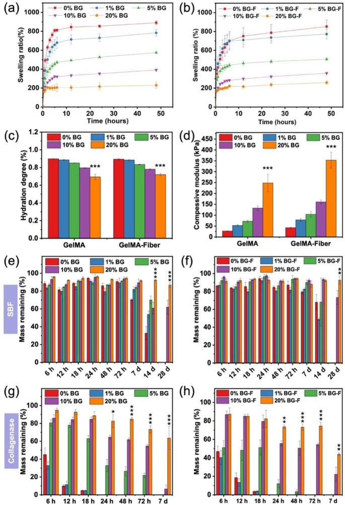
The GelMA/BG-Fiber composite hydrogels, containing various concentrations of BG (0, 1, 5, 10, 20% w/v), with or with out PCL@GelMA coaxial nanofibers, had been efficiently synthesized and may be crosslinked utilizing UV mild. Determine 1 exhibits that the brief coaxial nanofibers with various concentrations of BG had been efficiently integrated whereas preserving the porous construction of the hydrogel. The porous construction of hydrogels is crucial for cell progress and angiogenesis [26]. The composite hydrogels exhibited sturdy swelling properties as a consequence of their extremely interconnected porous construction. The swelling of hydrogels performs a pivotal position within the therapeutic and regeneration of bone tissue because it facilitates the transportation of vitamins and the elimination of waste merchandise by means of diffusion [21]. The incorporation of BG considerably influences the swelling conduct of the hydrogels. The swelling traits of the hydrogels decreased with the rise in BG content material, which can be attributed to the electrostatic interplay between the negatively charged silicon hydroxyl (–Si–OH) teams on the floor of BG and the positively charged amino (–NH2) teams in GelMA [9], whereas the incorporation of coaxial fibers had no vital impact (Fig. 2a-b). The hydration charge of the composite hydrogel with 20% BG-Fiber was 72%, which was decrease than that of GelMA (89%) (Fig. 2c). The outcomes reveal that the composite hydrogels exhibit favorable swelling ratios, which point out their potential for environment friendly nutrient diffusion when transplanted into the physique for bone regeneration.
The impact of BG and coaxial fibers on composite hydrogels’ hydration diploma, mechanical, and physiological stability. (a–c) The swelling charge and hydration diploma had been weakened by including BG and coaxial fibers. (d) The mechanical property was enhanced by including BG and coaxial fibers. (e–f) Degradation behaviors of hydrogels in SBF confirmed sluggish weight reduction after 28 days soaking. (g–h) Degradation behaviors of hydrogels in collagenase answer decreased the degradation charge by including BG and coaxial fibers. (*p < 0.05, **p < 0.01, ***p < 0.001)
The mechanical properties of the hydrogel had been considerably enhanced by incorporating PCL@GelMA coaxial nanofibers and BG. The GelMA shell of the coaxial fiber exhibited a exceptional affinity with the encircling GelMA hydrogel, facilitating crosslinking beneath UV mild irradiation and thereby augmenting the general mechanical traits. Throughout compression efficiency testing, the composite hydrogel containing PCL@GelMA coaxial nanofibers exhibited rupture at a compression stage of 65%. Remarkably, upon completion of the take a look at, the composite hydrogel absolutely recovered its preliminary look, whereas the absence of PCL@GelMA coaxial nanofibers within the hydrogel resulted in irreversible fragmentation (Fig. S4a). The two% focus of PCL@GelMA coaxial nanofibers of the composite hydrogel, solely supplemented with PCL@GelMA coaxial nanofibers, exhibited a considerably greater worth in comparison with the content material of 0% and 1%, whereas no vital distinction was noticed compared to the three% content material (Fig. S4b). This end result was much like that of Qiu [20], and due to this fact, 2% PCL@GelMA coaxial nanofibers had been used within the subsequent experiments. The incorporation of PCL@GelMA coaxial nanofibers not solely enhances the mechanical properties of the hydrogel but in addition maintains the hydrogel form. The hydrogel exhibited a simultaneous improve in final stress and compressive modulus with the addition of BG (Fig. 2d and Fig. S5a-b). The GelMA-containing BG hydrogels exhibited rupture at roughly 60% compression, whereas the composite hydrogel containing PCL@GelMA coaxial nanofibers exhibited rupture at a compression stage of 65%. Notably, hydrogels with a BG content material of 20% exhibited irrecoverable deformation when subjected to flat compression. Mechanical properties play a vital position within the improvement of bone regeneration supplies. Easy hydrogels possess restricted mechanical power and, due to this fact, necessitate the incorporation of PCL@GelMA coaxial nanofibers and BG to boost the general robustness. After incorporating these constituents, composite hydrogels retain their injectability and exhibit the potential for mending irregular bone defects.
The degradation properties of composite hydrogels are essential, and the optimum degradation situation is the synchronization of hydrogel degradation and bone progress [27]. The degradation of composite hydrogels was assessed throughout a 28-day incubation interval in SBF, approximating the length required for bone therapeutic and reworking following fracture [21]. In SBF answer, the composite hydrogels containing 0%, 1%, and 5% BG exhibited vital degradation inside two weeks and full degradation by 4 weeks, whereas the hydrogels with 10% and 20% BG content material demonstrated partial degradation (Fig. 2e-f). Collagenase-mediated degradation was fast and full inside 24 h for hydrogels with 0%, 1%, and 5% BG content material. Hydrogels containing 20% BG additionally exhibited vital degradation, reaching roughly 63% and 43% (fiber-containing) after 7 days (Fig. 2g-h). The addition of BG successfully slowed down the degradation course of, probably because of the sturdy interplay between BG and GelMA hindering the penetration of the collagenase answer. Nonetheless, incorporating coaxial fibers didn’t considerably hinder the degradation, probably because of the simultaneous degradation of the GelMA part within the shell and the hydrogel. The burden of the composite hydrogels exhibited fluctuations in the course of the degradation measurements, which might be attributed to the hydrogel degradation together with the deposition of phosphates, which led to a rise within the weight of the pattern. It may be inferred that the noticed mass change is primarily attributed to the preliminary dissolution of the BG and the next formation of mineralized crystals [28].
For the phosphate formation potential take a look at, the composite hydrogels of every group had been immersed in SBF for 7 days. Subsequently, the collected pattern sections had been examined utilizing SEM. Particles had been noticed on the floor of the composite hydrogels, and EDS evaluation revealed that these particles had been enriched in calcium (Ca) and phosphorus (P) (Fig. S6a-b), that are the primary parts of bones. The composite hydrogel’s Ca/P ratio of the floor components was 1.56 ± 0.03, which resembles that in synthetic bone (starting from 1.50 to 1.67) [29]. The deposition of phosphate crystals on the floor regularly will increase with rising BG content material (Fig. S6c), which is also seen on the floor of the fibers (Fig. 3). Within the presence of SBF, the composite hydrogels exhibited fast launch of calcium and phosphorus ions, forming calcium phosphate crystals on their floor [30]. Incorporating coaxial fibers additionally facilitates the nucleation and progress of phosphate crystals, enhancing their crystallization capability [31]. In conclusion, incorporating BG and coaxial fibers considerably mitigated swelling and hydration charge, decelerated degradation kinetics, and augmented mechanical properties and mineralization formation potential of the composite hydrogels.
The inorganic parts in bone biomaterials analysis embody BG, β-tricalcium phosphate, and hydroxyapatite [32,33,34]. After implantation into broken bones, BG is reabsorbed and built-in with the bone by forming a layer of apatite on its floor [35]. Though numerous polymers have been utilized in growing bone biomaterials, deciding on polymers that intently emulate the pure bone microenvironment is crucial. A number of research have utilized each artificial and pure polymers, comparable to chitosan, gelatin, and sodium alginate, to manufacture bone biomaterials containing BG [36,37,38,39]. The choice of gelatin, a pure polymer, is extremely advantageous for establishing an setting that intently emulates the first natural constituents of endogenous bones as a consequence of its hydrolyzed type derived from sort I collagen current within the skeletal system. Collagen can improve the metabolic exercise of osteoblasts, thereby selling osteogenesis, suppressing irritation, inducing chondrogenesis, and bettering bone mineral density [40]. In comparison with gelatin, GelMA exhibited the first benefit of a slower degradation charge and enhanced mechanical power. As well as, the interplay between cells and the ECM is regulated by arginine-glycine-aspartic acid sequences inside the GelMA construction, which function recognition websites for adhesive proteins that facilitate cell adhesion [41]. In abstract, GelMA/BG-Fiber composite hydrogel is a wonderful biomaterial that may successfully mimic the pure organic microenvironment of bone and has the potential to advertise bone restore and regeneration.
Impact of composite hydrogels on cell behaviors in vitro
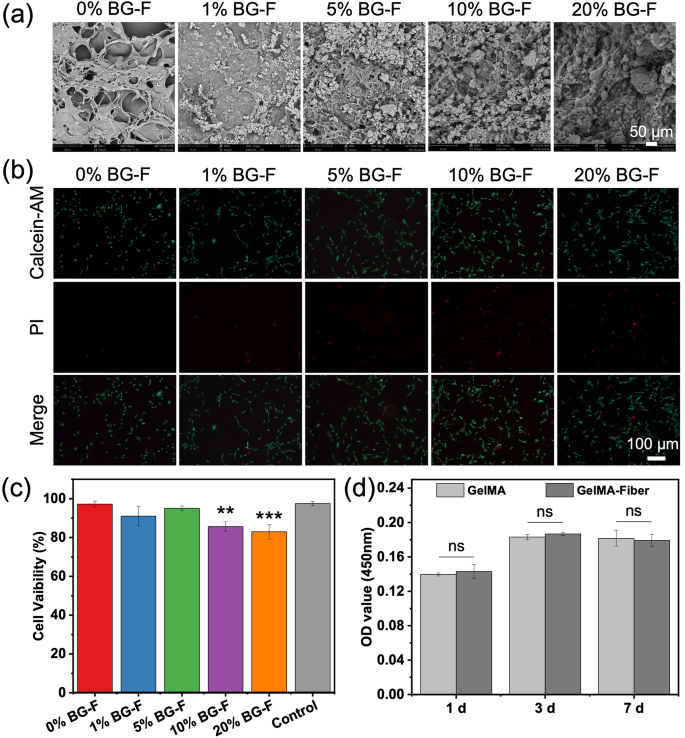
The interplay between cells and hydrogels is crucial. Upon examination of the cells adhered to the floor of the composite hydrogels, it was noticed that they exhibited sturdy adhesion, with even crystalline formations rising on their surfaces in the course of the sustained launch of ions from the BG (Fig. 4a). This phenomenon grew to become extra pronounced with rising BG content material. We initially investigated the impact of fiber supplementation on cell proliferation and noticed no statistically vital variations (Fig. 4d). The biocompatibility of various composite hydrogels was assessed utilizing Calcein-AM/PI (dwell/useless) staining. It was noticed that a rise within the content material of BG resulted in a better variety of useless cells, and 83% of the cells survived within the 20% BG of composite hydrogel within the extraction answer (Fig. 4b-c). We attribute these outcomes to crystal progress. The incorporation of BG has been proven to launch a big amount of ions, leading to an preliminary alkaline pH within the answer, which subsequently impacts cell proliferation [42]. The expansion and proliferation of stem cells are additionally influenced by the content material of BG in answer, with cell progress being stimulated solely at an acceptable focus [43]. Subsequently, on this experiment, we hypothesize that the event of crystals, adjustments in answer pH, and BG content material within the answer exert an inhibitory impact on cell proliferation.
The analysis of biocompatibility of composite hydrogels. (a) SEM photos of MC3T3-E1 on composite hydrogels at day 3. (b) Calcein-AM/PI (dwell/useless) staining of various composite hydrogels. (c) Outcomes of Calcein-AM/PI (dwell/useless) staining by counting the variety of dwell cells. (d) The CCK8 assay confirmed that the cell progress was inhibited with the rise in BG. (*p < 0.05, ***p < 0.001)
Angiogenesis and osteogenesis are important for bone regeneration in tissue engineering scaffolds. The affect of hydrogels with various BG contents on HUVEC tube formation assays was assessed. The composite hydrogel containing 20% BG exhibited essentially the most intensive tube community formation, steady tube partitions, and essentially the most vital variety of assembly factors, segments, and branches (Fig. 5a-d). Determine 5e exhibits cell migration photos of HUVECs after culturing with the extraction answer of the composite hydrogels for twenty-four and 48 h. The corresponding quantitative migration areas stuffed by HUVECs are proven in Fig. 5f-g. The composite hydrogel containing 20% BG exhibited bigger migration areas stuffed by HUVECs than the opposite teams. To evaluate the differentiating impact of hydrogels on cells, we remoted and characterised BMSCs primarily based on their multipotent differentiation potential and cell surface-specific markers (Fig. S7) after which cultured BMSCs within the extraction answer of composite hydrogels. By evaluating the alizarin crimson staining of BMSCs cultured for 14 days, it was noticed that the world of optimistic staining was considerably elevated within the composite hydrogels containing 10% (9.7%) and 20% BG (12.8%) (Fig. 6a-b). Quantitative evaluation of ALP confirmed that at 7 days, ALP expression was greater within the composite hydrogel containing 1% BG (0.24 U/L), whereas at 14 days, ALP expression was considerably elevated within the composite hydrogel containing 20% BG (0.27 U/L) (Fig. 6c). Relating to gene expression analyzed by qPCR, the inclusion of 5%, 10%, and 20% BG hydrogels considerably enhanced the expression of OPN when BMSCs had been cultured for 7 days. Moreover, after 14 days, incorporating a 20% BG composite hydrogel elevated the expression ranges of ALP (9.1 instances), RUNX2 (2.7 instances), and OPN (2.3 instances) in BMSCs (Fig. 6e-f). ALP is an early indicator of osteogenesis and intently correlates with the biomineralization course of. RUNX2 and OPN are essential ECM proteins that serve twin features, enjoying a pivotal position within the survival of osteoclasts in addition to bone mineralization [44]. The hydrogels containing 20% BG exhibited the best capability for mineralized crystal formation, which can transiently impede cell proliferation however in the end promote osteogenic differentiation. Consequently, this specific group was chosen for in vivo experimentation. Subsequently, including bioglass releases numerous ions, comparable to calcium, phosphorus, and silicon ions, and these can produce phosphate crystals when in touch with physique fluids. Such crystals have a excessive hardness, which might represent each the bone mechanism and the sign for osteogenic differentiation that may be generated for stem cells, selling the expression of osteogenesis-related genes comparable to ALP, RUNX2, OPN, which is conducive to the differentiation of cells to osteoblasts.
Impact of scaffolds on tube formation of angiogenesis. (a) Tube formation photos of HUVECs. (b) Variety of junctions, (c) whole branching size, and (d) whole phase size per area of view. (e) The pictures of HUVECs cultures for twenty-four and 48 h and (f, g) quantitative statistics of cell migration charges in cell scratch assay (n = 3). (*p < 0.05, **p < 0.01, ***p < 0.001)
The osteogenic differentiation detected by alizarin crimson staining, ALP quantification, and qPCR. (a–b) Alizarin crimson staining after cultured BMSCs with the composite hydrogel extraction answer for 14 days. The ten% and 20% BG composite hydrogels can considerably improve the optimistic staining space. (c) Quantitative evaluation of ALP exhibits that the expression of ALP within the composite hydrogel with 1% BG content material was greater at 7 days, whereas the composite hydrogel with 20% BG content material considerably promoted ALP expression at 14 days. (d–e) qPCR outcomes detecting the osteogenic gene (ALP, RUNX2, and OPN) expression of BMSCs cultured with completely different composite hydrogels. (*p < 0.05, ***p < 0.001)
Bone regeneration promoted by composite hydrogels in vivo
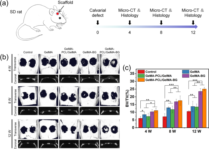
To guage the osteogenic potential of the composite hydrogels in vivo, we initially implanted the lyophilized hydrogels into the subcutaneous tissue of rats. Subsequently, we obtained samples for Micro-CT evaluation after 2 and 4 weeks. In comparison with hydrogels with out fibers, composite hydrogels containing fibers confirmed considerably enhanced mineralization after 2 and 4 weeks in vivo (Fig. S8). The fibers might supply further attachment websites to type mineralized crystals, selling crystal nucleation and subsequent crystal progress. Subsequently, a rat cranium defect mannequin was established, and the composite hydrogel was injected into the defect web site, adopted by UV mild curing for 10 s. The samples had been collected and analyzed by Micro-CT at 4, 8, and 12 weeks. Subsequently, the samples had been decalcified, sectioned, and stained with H&E, Masson’s trichrome stain, and immunohistochemical staining (Fig. 7a). The 20% BG composite hydrogel demonstrated vital osteogenesis in Micro-CT detection in comparison with the opposite teams in any respect three-time factors (Fig. 7b). Furthermore, though the presence of coaxial fibers didn’t exert a big impact on bone regeneration, it was noticed that the bone tended to be situated extra centrally inside the cranium defect. The quantitative bone quantity/tissue quantity (BV/TV) information was computed primarily based on the 3D Micro-CT information. At 12 weeks, the 20% BG composite hydrogels repaired roughly 25% and 23% of the bone defects, which was a lot greater than the management group (10%) (Fig. 7c). This discovering means that fibers might function nucleation websites for crystal formation, facilitating and enhancing bone regeneration. At present, the prominence of 3D printing know-how is driving developments in bone formation, however it’s difficult to use to restore irregular defects [45, 46]. The power of our examine lies in its potential to cater to numerous software eventualities involving irregular defects whereas being simply implementable.
The animal experiment patterns and evaluation of Micro-CT outcomes. (a) The cranium defect mannequin of 6 mm diameter was established, and the samples had been analyzed at 4, 8, and 12 weeks. (b–c) Micro-CT outcomes confirmed that including BG and coaxial fiber induced extra bone regeneration. (*p < 0.05, **p < 0.01, ***p < 0.001)
The outcomes of H&E and Masson’s trichrome staining of every group are offered in Fig. 8. The GelMA group was nonetheless not utterly degraded at 4 weeks. Nonetheless, upon the addition of BG, a considerable formation of blood tissue and bone-like matrix was noticed, accompanied by an enhanced infiltration of lymphocytes. At 8 weeks, the BG group exhibited extra multinucleated osteoclasts, whereas lymphocytes remained plentiful. By 12 weeks, lymphocyte ranges began to say no, accompanied by a rise within the amount and dimension of the osteoid matrix and a big discount within the osteoclast inhabitants. As proven in Fig. 9a-b, hydrogels (together with BG and coaxial fibers) dramatically elevated the expression of OPN in immunohistochemical staining in any respect durations. Particularly at 12 weeks, OPN expression within the GelMA-PCL@GelMA-BG group was 5 instances greater than within the management group. CD31, a marker of nascent endothelial cells, is usually employed for neonatal microvessel quantification to evaluate the angiogenesis of implanted supplies [47, 48]. In immunohistochemical staining for CD31, the GelMA-PCL@GelMA-BG group had extra CD31 expression and extra vascular tissue manufacturing at 8 and 12 weeks (Fig. 9c-d). The noticed outcomes could also be attributed to the synergistic interplay between the hydrogel matrix and the particles launched from BG. Within the GelMA-BG group, a considerable quantity of CD31-labeled cavity formation was noticed, which was rare within the GelMA-PCL@GelMA-BG group. Consequently, these findings recommend that together with coaxial fibers successfully restricts cavity formation. It has been demonstrated that silicates can upregulate nitric oxide synthase, thereby inducing angiogenesis [49]. Calcium ions additionally facilitate angiogenesis, thereby augmenting the secretion of vascular-related cytokines and selling endothelial cell adhesion [50]. BG, which releases calcium ions, might facilitate this course of. Furthermore, blood vessels can ship vitamins, bioactive elements, and osteogenesis-related cells to advertise new bone formation and transport metabolic waste or poisonous merchandise for accelerated restore. In conclusion, the novel benefits of the GelMA/BG-Fiber composite hydrogel enhanced biocompatibility and improved osteogenic and angiogenic marker expression, that are essential for selling bone regeneration.
Immunohistochemical staining of the samples. (a–b) Immunohistochemical staining of osteoblastic marker (OPN) exhibits that the addition of BG and coaxial fibers promotes the expression of OPN. (c–d) Immunohistochemical staining of angiogenic marker (CD 31) exhibits that including BG and coaxial fibers promotes the expression of CD 31 at 8 and 12 weeks. (*p < 0.05, **p < 0.01, ***p < 0.001)
However the preliminary findings of the current examine, sure limitations had been recognized. As an illustration, the composite hydrogel shows insufficient power, rendering it incapable of offering sufficient help for the bone defect on the weight-bearing web site. Consequently, it may well solely be employed as an auxiliary measure subsequent to inside fixation. Secondly, the hydrogel is of low viscosity, which carries the chance of detachment from the bone defect web site. It’s our intention to additional enhance the hydrogel composite so as to facilitate osteogenesis and vascularisation, which might be extra pertinent to the context of bulk bone defect surgical procedure.


