Albumin-seeking NIR dyes covalently mix with albumin and improve brightness
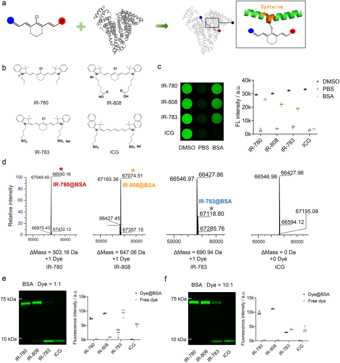
We proposed a chemoselective albumin-seeking technique for NIR dyes binding to albumin in vivo. Earlier research have proved the covalent bonding between chlorine-containing (Cl-containing) cyanine dyes and albumin [27]. IR-780, IR-808 and IR-783 have been cyanine dyes possessing chlorocyclohexene that would first enter the albumin pocket by supramolecular interactions and underwent covalent binding by way of a nucleophilic substitution response between the -SH group in albumin and the Cl-C bond in these dyes [27, 28]. Whereas the Cl-free dye (ICG) sure albumin solely by non-covalent supramolecular interactions [29]. Covalent binding of Cl-containing cyanine dyes and bovine serum albumin (BSA) enhances the steadiness and brightness of free cyanine dyes (Fig. 1a) [13, 30, 31]. We chosen the Cl-containing NIR cyanine dyes that may covalently bind to albumin with optimum albumin-binding talents of IR-780 > IR-808 > IR-783, and ICG as a comparability which not covalently bind to albumin (Fig. 1b) [27]. First, we in contrast the brightness traits of the 4 probes in vitro binding to albumin. The dyes exhibited the best and lowest brightness in DMSO and PBS, respectively. Dyes in BSA have been incubated at a 1:1 molar ratio at 60 °C for two h. The absorption and fluorescence spectra confirmed that the free dyes and Dye@BSA in PBS exhibited absorption peaks at 779–799 nm with NIR-II tail emission (Fig. S1). IR-780, IR-808, and IR-783 have been related to the next brightness forming Dye@BSA than free dyes in PBS (Fig. 1c). As well as, in contrast with dyes in PBS, Dye@BSA has greater NIR-II quantum yield (Fig. S2, S3, Desk S1). Subsequent, the binding proportions of the 4 dyes to BSA have been analyzed. Mass spectrometry illustrated that IR-780, IR-808, IR-783 sure to BSA in a 1:1 ratio (Fig. 1d). The bio-layer interferometry (BLI) experiments quantitatively in contrast binding affinity of IR-780 (OkD = 2.80E + 04 nM) > IR-808 (OkD = 9.50E + 04 nM) > IR-783 (OkD = 4.30E + 05 nM) > ICG (OkD =8.10E + 05 nM) (Fig. S4, Desk S2). Gel electrophoresis confirmed that IR-780 sure nearly fully to BSA, IR-808 sure largely to BSA with a small fraction of free IR-808, IR-783 sure solely partially to BSA, and ICG didn’t kind any secure complicated with BSA (Fig. 1e). As albumin is essentially the most considerable protein within the blood, and former research have proven that growing the BSA: dye ratio enhances the binding proportion [27, 32, 33]. Subsequently, we additional incubated BSA with dyes at a ratio of 10:1. The gel electrophoresis revealed an enhanced binding of Cl-containing cyanine dyes to BSA, with IR-780 and IR-808 binding nearly fully to BSA and IR-783 exhibiting an elevated binding to BSA, whereas ICG binding proportion remained unchanged (Fig. 1f).
Chlorine-containing albumin-seeking dyes covalently bind to albumin and improve brightness. (a) Schematic diagram of the covalent binding web site of chlorine-containing cyanine dye and albumin. (b) Structural system of IR-780, IR-808, IR-783, and ICG. (c) Brightness of 4 dyes (50 nM) in DMSO, PBS, and BSA with incubating at 60 ºC of 1:1 molar ratio (imply ± SEM, n = 3). (d) Mass spectrometric evaluation of 4 dyes in BSA with 1:1 response molar ratio at 37 ºC for two h. (e–f) Gel electrophoretic evaluation of fluorescence depth of 4 dyes in BSA at 1:1 and 10:1 response molar ratio at 37 ºC for two h (imply ± SEM, n = 3)
Albumin-seeking dyes extremely affinity and selectively bind to albumin in situ with enhanced brightness and circulation time
Earlier than making use of albumin-seeking dyes to stroke imaging, we noticed the systemic metabolism and circulation of the 4 dyes in mice. After intravenous injection, IR-780 and IR-808 gathered within the liver after which distributed all through the entire physique sustaining excessive brightness for about 72 h with an extended metabolism time. In distinction, IR-783 and ICG gathered within the liver and cleared quickly from the physique inside 24 h (Fig. S5).
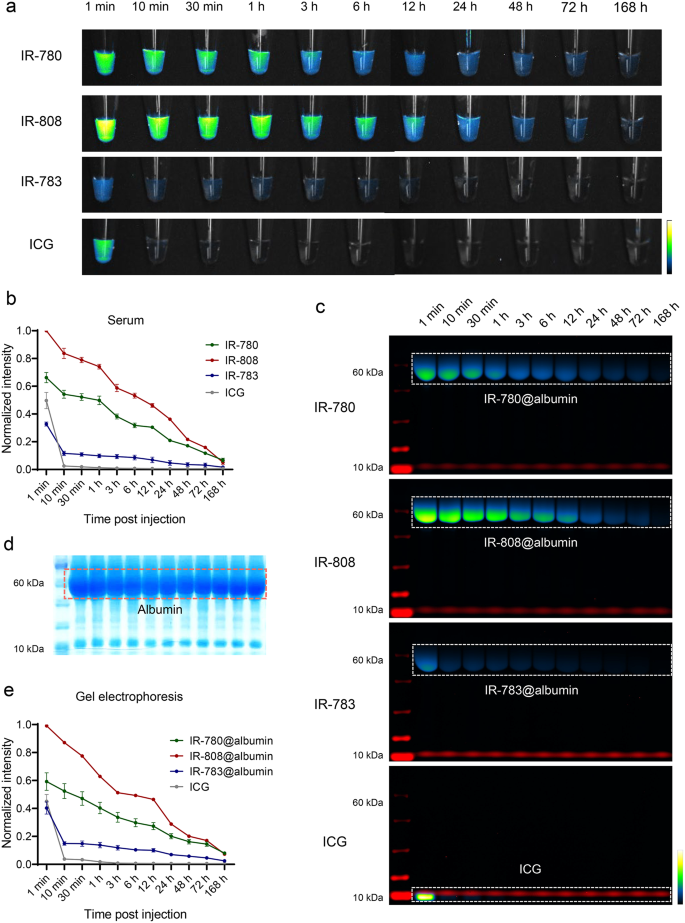
To confirm the metabolism of dyes within the circulation and the formation of dye@albumin complexes in vivo, blood samples have been collected from mice intravenously injected with the identical dose of the 4 dyes. The serum was separated and subjected to gel electrophoresis, adopted by NIR fluorescence imaging and gel staining with Coomassie Sensible Blue. IR-780 and IR-808 maintained greater brightness within the circulation for an extended interval in comparison with IR-783 and ICG, which regularly decreased inside 7 days after the tail venous injection of the probes (Fig. 2a, b). In distinction to the outcomes of brightness in vitro binding to albumin, IR-808 exhibited the best brightness in circulation, adopted by IR-780. It could be attributed to the hydrophobicity of IR-780, leading to self-assembly and decrease brightness when injected into the mice than IR-808 [34]. The serum gel electrophoresis outcomes confirmed that IR-780, IR-808, and IR-783 shaped dye@albumin complexes of roughly 66 kDa in situ, whereas ICG remained in its free kind and was quickly metabolized (Fig. 2c). Determine 2d reveals the band patterns of assorted proteins within the serum collected at totally different time factors. These outcomes indicated the excessive selectivity and stability of IR-780, IR-808, and IR-783 binding to albumin. According to the serum fluorescence brightness outcomes, the IR-808@albumin band was the brightest and the IR-780@albumin band was the second brightest. Solely a portion of IR-783 shaped complexes with albumin within the bloodstream, whereas the unbound portion of the dye was shortly metabolized and excreted. ICG didn’t bind to endogenous albumin and exhibited the quickest metabolism, reaching its highest brightness after 1 min, which decreased quickly 10 min post-administration (Fig. 2e). These outcomes illustrate that albumin-seeking probes can selectively and stably bind albumin in vivo with growing brightness and circulation time.
Comparability of circulation metabolic habits of albumin-seeking dyes in vivo. (a) Serum was collected at time factors after tail vein injection (50 µM, 60 µL) of IR-780, IR-808, IR-783 and ICG and imaged below the NIR-II window (80 ms, 1100 LP). (b) Normalized fluorescence depth of serum after injection of dyes (imply ± SEM, n = 3 for every group). (c) Gel electrophoresis evaluation of dyes and albumin binding modified over time in mice serum. (d) Coomassie sensible blue confirmed protein distribution of serum after gel electrophoresis. (e) Normalized fluorescence depth of albumin complicated band of IR-780@albumin, IR-808@albumin, and IR-783@albumin and free ICG (imply ± SEM, n = 3 for every group)
Albumin-seeking dyes monitoring BBB disruption in stroke mice in real-time
Albumin is often scarce within the mind tissue. Nonetheless, blood albumin enters the mind by the broken BBB post-stroke [35, 36]. Subsequently, the albumin in mind serves as a biomarker for BBB damage [25, 26]. Though the dye-albumin complicated enhanced the brightness and circulation time of free dye, introducing exogenous albumin could set off immune or inflammatory reactions [37]. Subsequently, the probe that may bind to endogenous albumin in situ could supply protected imaging and good capabilities for monitoring BBB disruption.
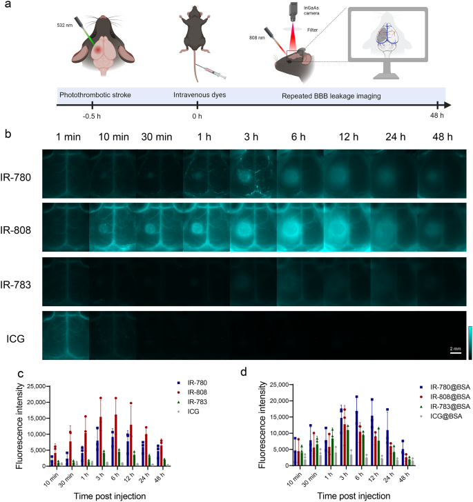
We explored albumin-seeking dyes for visualization of BBB disruption after stroke in a photothrombotic stroke (PTS) mannequin with infarcts within the left cerebral cortex of mice. At 30 min submit stroke, IR-780, IR-808, IR-783, and ICG have been intravenously injected on the similar dose respectively, and real-time dynamic NIR-II imaging was carried out with out the necessity for craniotomy (Fig. 3a). As proven in Fig. 3b, cerebral vessels have been visualized however no fluorophore leakage into the stroke area at 1 min after probes injection. Fluorescence indicators regularly gathered within the stroke space within the IR-780 and IR-808 teams with leakage noticed as early as 10 min, indicating the buildup within the mind parenchyma by the broken BBB. IR-808 exhibited a brighter imaging than IR-780, per the relative brightness of the serum. IR-783 have weaker brightness than IR-780 and IR-808, and confirmed a low fluorescence sign at 1 min. Because of the comparatively weak binding affinity of IR-783 with albumin, free IR-783 cleared quickly from the physique [27]. Subsequently, unbound IR-783 was quickly metabolized and thus confirmed decrease fluorescence indicators in cerebral vascular at 10 min / 30 min / 1 h than these at 1 min. After an extended interval of injection into the blood circulation, IR-783 might bind to albumin and tremendously improve its fluorescence depth, thus exhibiting the next fluorescence sign at 3 h than that at 10 min / 30 min / 1 h and accumulation of fluorescence indicators within the BBB leakage area was noticed however with decrease brightness than IR-780 and IR-808. ICG confirmed greater brightness within the blood vessels 1 min after injection however was quickly metabolized inside 10 min, with minimal sign accumulation within the stroke space. To attenuate the affect of brightness within the blood vessels, we quantified the fluorescence sign within the BBB disruption space by calculating the distinction between the sign within the BBB disruption space and corresponding sign within the wholesome contralateral space (Fig. 3c). IR-780, IR-808, and IR-783 confirmed the biggest distinction within the accumulation of fluorescence indicators at 6 h, whereas ICG exhibited minimal sign accumulation within the mind tissue, which regularly decreased after peaking at 1 h.
As well as, we validated the imaging capability of the BBB utilizing the dye@BSA complicated synthesized in vitro (Fig. S6a). IR-780@BSA, IR-808@BSA, and IR-783@BSA complexes exhibited comparable brightness and circulation instances in monitoring BBB disruption. ICG@BSA exhibited a habits just like free ICG. The brightness of the BBB disruption space was noticed to be IR-780@BSA > IR-808@BSA > IR-783@BSA (Fig. 3d). Though IR-780 demonstrated the strongest binding capability to albumin in vitro, it didn’t obtain comparable results as IR-780@BSA complicated in vivo, probably due to its hydrophobicity resulting in self-assembly, and inadequate binding in vivo [38]. In distinction, IR-808 has comparable results because the IR-808@BSA complicated, indicating that IR-808 can function a super albumin-seeking probe for monitoring BBB disruption after stroke with out the necessity for exogenous albumin, thus providing higher biocompatibility. To verify that BBB imaging utilizing IR-808 depends on its in vivo binding to albumin, we used an albumin-escaping probe, IR-808AC [39], which clearly visualized blood vessels however didn’t accumulate nor present BBB disruption and was fully metabolized from the blood vessels inside 10 min (Fig. S6b).
Albumin-seeking dyes real-time monitoring BBB disruption in mice with stroke. (a) Schematic diagram of photothrombotic stroke mannequin and NIR-II imaging for BBB disruption monitoring post-stroke (Created with BioRender.com). (b) NIR-II dynamic imaging (40 ms, 1100 LP) recorded at indicated time factors post-injection of stroke mice with IR-780, IR-808, IR-783, and ICG (50 µM, 60 µL). (c–d) Distinction in fluorescence sign depth on the left and proper sides post-injection of free dyes and dye@BSA (imply ± SEM, n = 3 for every group)
The albumin-seeking dyes permits for quick evaluation, multi-timepoint monitoring after stroke and a real-time, high-resolution visualization of BBB disruption. Among the many 4 dyes examined, IR-808 exhibited essentially the most exceptional visualization of BBB disruption. Subsequently, IR-808 is a promising probe for correct evaluation of stroke damage and we performed detailed imaging utilizing IR-808 in our subsequent investigations.
IR-808 permits analysis of BBB damage in strokes with varied severities
We first displayed the imaging of IR-808 monitoring BBB leakage in NIR sub-windows. Fig. S7a presents the imaging outcomes of IR-808 in NIR-I window (850–900 nm) and three sub-NIR-II home windows (> 1100 nm, > 1200 nm, and > 1300 nm). IR-808 might present enough brightness below the longer NIR-II sub-window by growing the publicity time and improves signal-to-background ratio (SBR) in contrast with in NIR-I window (Fig. S7b). We subsequent noticed adjustments in cerebral perfusion and IR-808 fluorescence sign accumulation in acute-phase of stroke. Dynamic steady imaging of cerebral arteries and veins within the mice was noticed 2.8 s submit IR-808 injection. In contrast with wholesome facet, a discount in cerebral perfusion was noticed within the infarct space of stroke mice (Fig. S8a, b). Leakage of fluorescent sign was noticed within the stroke space 5 min after injection of IR-808, and fluorescent sign accumulation elevated inside 1 h (Fig. S8c, d).
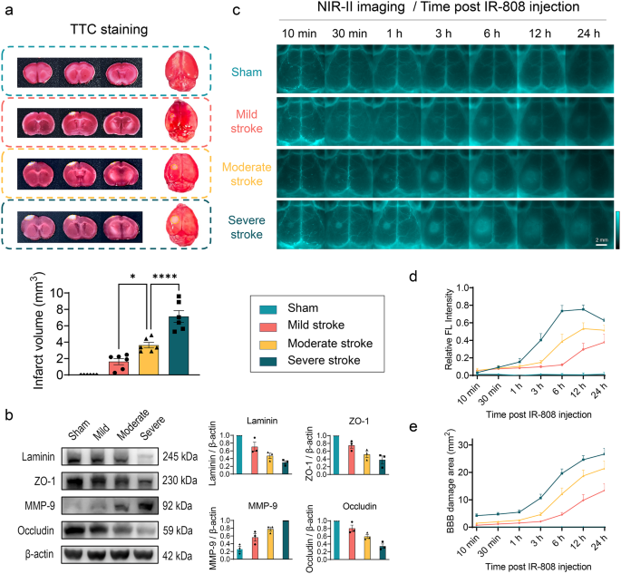
We additional explored the effectivity of IR-808 to detect and distinguish BBB damage of sham mice and three severity ranges of stroke. As described within the strategies, the severity of stroke damage was graded into three teams utilizing the PTS mannequin [40]. TTC outcomes confirmed a progressive growing infarct quantity at 24 h after stroke in gentle, reasonable and extreme stroke teams (Fig. 4a). Western blotting evaluation of protein expression in mind tissue confirmed a gradual lower in tight junction-related proteins (ZO-1, occludin) and basement membrane-related protein (laminin) and a rise in matrix metalloproteinase (MMP-9) ranges within the sham, gentle, reasonable, and extreme stroke teams (Fig. 4b), indicating an elevated diploma of BBB disruption. NIR-II imaging reveals the fluorescence indicators have been comparable in each side and didn’t leak by the intact BBB within the sham group. In stroke teams, fluorophores leakage initially within the infarct core and regularly accumulates within the peri-infarct tissue as stroke time progressing, indicating dynamic aggregation by leakage BBB into mind tissue (Fig. 4c). Relative fluorescence depth of the affected facet elevated progressively after stroke. Within the reasonable to extreme stroke, the relative fluorescence depth peaked at 12 h, whereas within the gentle stroke it elevated till the 24-h peak (Fig. 4d). The BBB leakage space elevated over time in stroke teams and was bigger within the worse stroke teams (Fig. 4e). As well as, we analyzed statistical variations in BBB disruption between stroke severity at time factors. The relative fluorescence depth was considerably greater within the extra severity stroke teams at 6 h (P < 0.01) and 12 h (P < 0.05) (Fig. S9a). The BBB injury space was considerably totally different at 6 h between the three stroke teams (P < 0.01) and was considerably decrease in gentle stroke than in moderate-to-severe strokes at 12 h (P < 0.01) (Fig. S9b). At 24 h, relative fluorescence depth and injury space have been considerably greater in extreme stroke than gentle stroke (P < 0.05). These outcomes point out that IR-808 permits dynamic evaluation of strokes, in addition to good variations in leakage space and relative fluorescence depth measured by IR-808 in varied severity strokes.
IR-808 dynamic monitoring of BBB disruption with various severity. (a) Coronal slices and whole-brain TTC staining in sham, gentle, reasonable, extreme stroke teams and statistical evaluation of infarct volumes quantified in slices (imply ± SEM, n = 6 for every group). (b) Western blotting evaluation of BBB damage protein expression within the brains of mice with stroke with incremental severity (imply ± SEM, n = 3 for every group). (c) IR-808 NIR-II imaging for in vivo multi-timepoint monitoring of BBB disruption in strokes of various severity (40 ms, 1100 LP). (d) Quantification of the relative FL depth (Ratio of fluorescence depth of affected facet/wholesome side-1) at time post-injection (n = 6 for every group). (e) Quantification of the world of fluorescence sign accumulation within the BBB injury space at time post-injection (n = 6 for every group). *P < 0.05, ****P < 0.0001
BBB disruption evaluated by IR-808 correlates properly with Evans blue measurement
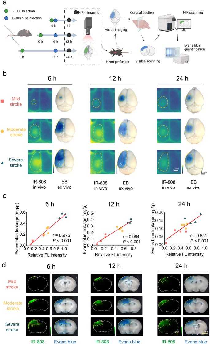
Presently, BBB disruption in experimental animals is especially assessed by intravenous injection of Evans blue (EB), adopted by ex vivo imaging and quantification [41]. To confirm the reliability of the IR-808 in vivo analysis of BBB damage, we carried out when it comes to each IR-808 NIR-II imaging in vivo and EB ex vivo measurement and quantification in sections at 6 h, 12 h, and 24 h, respectively. IR-808 leakage space and fluorescence depth in addition to EB leakage space and quantification have been analyzed in the identical mice (Fig. 5a).
Determine 5b current IR-808 in vivo and EB ex vivo imaging of gentle, reasonable, and extreme strokes at 6 h, 12 h, and 24 h post-stroke. The relative fluorescence depth of IR-808 correlated properly with EB quantification at 6 h (r = 0.975, P < 0.001), 12 h (r = 0.964, P < 0.001), and 24 h (r = 0.851, P < 0.001) (Fig. 5c). This means that the quantitative analysis of BBB damage through the use of IR-808 in vivo imaging is of excessive reliability. We additional analyzed the findings within the mind sections (Fig. 5d). EB leakage and accumulation of IR-808 fluorescence sign might be quantified in sections (Figs. S10, S11). The fluorescence depth quantification of IR-808 counted in sections correlated properly with the relative fluorescence depth in vivo (6 h: r = 0.921; 12 h: r = 0.857; 24 h: r = 0.879, all P < 0.001), demonstrating that in vivo NIR-II imaging has an excellent accuracy in reflecting IR-808 leakage into mind tissue (Fig. S12a). And the NIR fluorescence depth within the sections correlated with EB quantification validating the outcomes of in vivo imaging (Fig. S12b). Moreover, the dimensions ratio of BBB disruption in entire mind measured by IR-808 to EB at 6 h, 12 h and 24 h was 1.045 ± 0.039, 1.005 ± 0.047, and 1.396 ± 0.063, respectively, indicating that the IR-808 is dependable in assessing the world of leakage (Fig. S12c, d). Evaluating a number of time factors in vivo and ex vivo with EB, our outcomes exhibit the accuracy of IR-808 NIR-II imaging for repeat evaluation of BBB damage in vivo.
Correlation of IR-808 with Evans blue analysis of BBB damage. (a) Schematic diagram exhibiting the quantification of IR-808 and Evans blue (EB) on BBB disruption analysis in the identical mice (Created with BioRender.com). (b) IR-808 in vivo NIR-II imaging and its ex vivo seen EB imaging in mice with gentle, reasonable, and extreme stroke at 6 h, 12 h, and 24 h. Orange dotted line outlines BBB leakage areas. (c) Correlation of relative FL depth of IR-808 with Evans blue quantification at 6 h, 12 h, and 24 h (n = 12 every time level). (d) The corresponding photos of mind sections below NIR and visual scanning in panel b
As well as, to discover the correlation between BBB leakage assessed by IR-808 and remaining infarct quantity, we additionally assessed the correlation at 6 h, 12 h and 24 h of dynamic monitoring in varied severities stroke. The relative fluorescence depth at 6 h (r = 0.903, P < 0.001), 12 h (r = 0.825, P < 0.001), and 24 h (r = 0.659, P = 0.003) in addition to the world of BBB leakage at 6 h (r = 0.880, P < 0.001), 12 h (r = 0.785, P < 0.001), and 24 h (r = 0.779, P < 0.001), correlated with the ultimate infarct quantity (Fig. S13). These outcomes counsel that monitoring BBB disruption by IR-808 permits for early prediction of the ultimate infarct quantity throughout dynamic monitoring.
Twin-channel NIR-II dynamic imaging concurrently evaluates BBB damage and cerebral perfusion
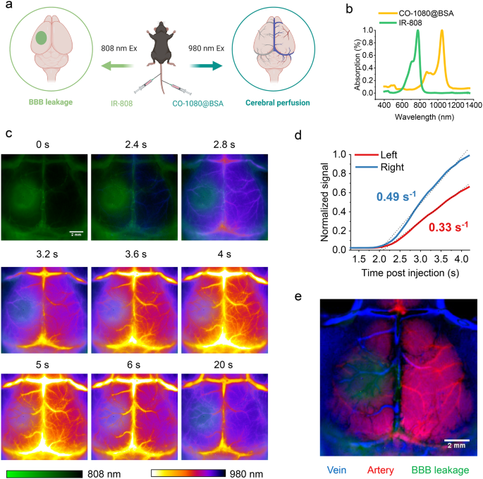
Mind neuropathology adjustments complicated after stroke, and adjustments in BBB injury typically accompanied with dynamic adjustments in cerebral perfusion. Thus, dual-channel NIR-II imaging for simultaneous analysis of BBB damage and cerebral perfusion has a excessive medical applicability. IR-808 below 808 nm excitation confirmed the world of BBB disruption. To evaluate cerebral perfusion whereas monitoring the BBB, we employed one other NIR-II probe, CO-1080@BSA, below 980 nm excitation and was intravenously injected at 6 h after stroke (Fig. 6a, b) [42]. Dynamic cerebrovascular photos have been captured starting with injection of CO-1080@BSA. NIR-II photos confirmed BBB leakage (808 nm, IR-808) and dynamic adjustments of the cerebrovascular arterial to venous part (980 nm, CO-1080@BSA) within the two NIR-II imaging channels (Fig. 6c). We quantified the dynamic fluorescence indicators of the cerebral blood vessels within the BBB leakage space and corresponding wholesome facet. In contrast with that within the wholesome facet, the cerebral perfusion was considerably diminished within the BBB leakage space (Fig. 6d). We carried out principal part evaluation (PCA) on a sequence of 40-frame dynamic photos to tell apart the arteries (crimson) and veins (blue) within the mouse mind vasculature and merged them with BBB leakage (inexperienced) (Fig. 6e). PCA revealed diminished arteries within the core of the ischemic space, whereas arterial vessels have been nonetheless current within the surrounding BBB damage space. The actual-time dual-channel NIR-II imaging system can observe adjustments in cerebral perfusion dynamics whereas evaluating BBB damage, which helps to perception the mechanism of neurological damage after stroke, and has good potential for in vivo and medical software.
Twin-channel NIR-II imaging of BBB disruption and cerebral perfusion after stroke. (a) Schematic diagram of dual-channel for BBB and cerebral perfusion imaging (Created with BioRender.com). (b) The absorption spectra of IR-808 and CO-1080@BSA. (c) Twin-channel photos of the BBB and cerebral vascular (100 ms, 1200 LP) after stroke at time submit CO-1080@BSA injection. IR-808 was used to guage BBB disruption utilizing 808 nm laser excitation, whereas CO-1080@BSA was used to dynamically consider the cerebral vascular utilizing 980 nm excitation at 6 h submit stroke. (d) Fluorescence depth (normalized to the right-side peak) versus time within the left (infarct facet) and proper (contralateral) sides of the mind of mice with stroke confirmed a big discount in perfusion within the stroke space. (e) Dynamic sequential photos of vessels with CO-1080@BSA have been analyzed by principal part evaluation and merged with photos of the BBB leakage space (inexperienced) to resolve arterial (crimson) and venous (blue) cerebral vessels
NIR-II BBB imaging supplies real-time evaluation of thrombolytic remedy efficacy and favorable biosafety
Thrombolytic remedy is a vital reperfusion remedy in acute ischemic stroke, however reperfusion could irritate BBB disruption [43, 44]. Subsequently, it’s obligatory to guage the BBB and cerebrovascular perfusion in actual time. We carried out NIR-II BBB imaging in a mouse thrombolytic remedy mannequin, reaching simultaneous dynamic evaluation of cerebrovascular perfusion and BBB damage earlier than and after thrombolysis (Fig. S14a). After stroke, center cerebral artery (MCA) occlusion and diminished perfusion on the affected facet have been noticed utilizing IR-808 at 808 nm channel (Fig. S14b). After thrombolysis, MCA revascularization and perfusion restoration have been noticed at 980 nm channel utilizing CO-1080@BSA (Fig. S14c). In the meantime, BBB damage might be evaluated earlier than and after thrombolysis in real-time, with the potential to information therapy (Fig. S14d-f). To additional confirm the universality of our technique, we efficiently carried out dynamic imaging of BBB disruption in a mouse everlasting center cerebral artery occlusion mannequin (Fig. S15) and a rat PTS mannequin (Fig. S16). These outcomes exhibit the potential extensive applicability of albumin-seeking probe mixed with NIR-II imaging for stroke BBB analysis.
Contemplating its potential medical purposes, we examined full metabolism in stroke mind tissue, impact on BBB restoration after stroke, and systemic security. Fluorescence indicators accumulating in stroke mind tissue have been fully metabolized 2 weeks after IR-808 injection (Fig. S17a-b). At 14 d, the consequences of the retention of IR-808 on mind tissue operate have been evaluated by behavioral checks and H&E staining. Behavioral checks outcomes confirmed no important distinction in unilateral sensorimotor dysfunction and cognitive operate between IR-808 group and PBS group (Fig. S17c). H&E staining confirmed the histopathological adjustments of peri-infarct tissue at 14 d after stroke was not considerably totally different between IR-808 group and PBS group (Fig. S17d). These outcomes point out that IR-808 retention didn’t impair mind tissue operate. As well as, BBB leakage was measured by EB in stroke mice injected with IR-808 or PBS at 7 d, 14 d, and 21 d after stroke, respectively. The outcomes confirmed no important distinction in EB leakage between the IR-808 and PBS teams, indicating that using IR-808 has no influence on BBB restore after stroke (Fig. S17e). As well as, serum from mice co-injected with IR-808 and CO-1080@BSA confirmed blood clearance below 808 nm and 980 nm excitation, respectively (Fig. S18a). Liver operate, kidney operate, and blood routine indices have been inside the regular vary in mice handled with IR-808 or IR-808 plus CO-1080@BSA (Fig. S18b). H&E staining confirmed no important histological injury to the center, liver, spleen, lungs, kidneys, or mind in comparison with the PBS group (Fig. S18c). These findings counsel that IR-808 and the co-injection of IR-808 plus CO-1080@BSA was protected and promising for medical translation to in vivo NIR-II imaging of stroke.


