Preparation and focus efficiency of SAPs with ionic community
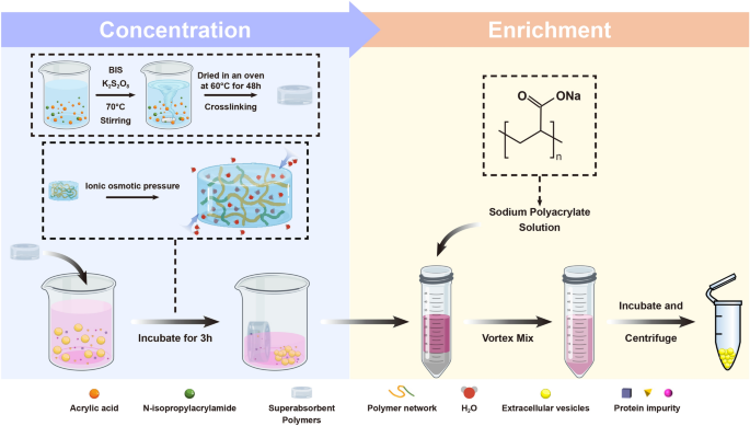
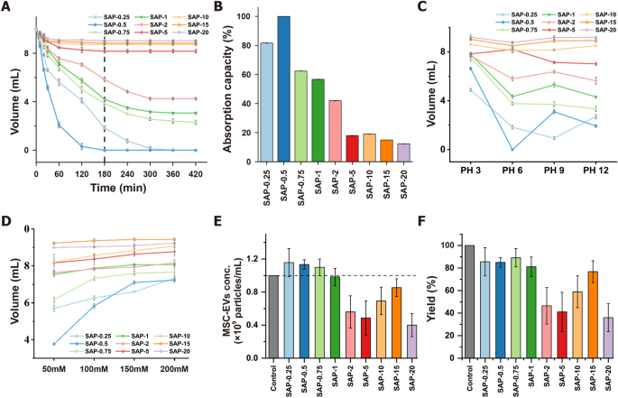
Quantity discount of CCM is important for the large-scale isolation of EVs. SAPs can focus CCM by ionic osmotic pressure-driven water absorption [20]. Water absorption effectivity of SAPs is influenced by the scale of water channels fashioned by ion networks, which in flip is affected by salt and pH. On this research, SAPs with gradient crosslinking levels and two monomers had been ready to optimize water absorption (Scheme 1, Desk S1). The SAPs had been categorized into low (SAP-0.25, SAP-0.5, and SAP-0.75), medium (SAP-1, SAP-2, and SAP-5), and excessive (SAP-10, SAP-15, and SAP-20) crosslinked teams based mostly on their completely different crosslinking levels. The adsorption conduct of SAPs was investigated by assessing their water quantity discount properties. The water quantity decreased with growing incubation time and reached a minimal after 180 min for various crosslinking levels (Fig. 1A). For instance, utilizing 10 mg/mL of SAP-0.75, the common water absorption fee was 34.07 µL/min within the first 180 min, and between 180 and 420 min, it slowed to six.53 µL/min diminished to 38.67% underneath these situations. The saturation water absorption capability of SAP-0.75 was 13.62 occasions that of the business SAPs [20]. As proven in Fig. 1B, the water absorption capability of SAPs decreased considerably with growing crosslinking levels, the place low crosslinked SAPs exhibited the best adsorption capability.
Characterization of superabsorbent polymers (SAPs) for concentrating extracellular vesicles (EVs) through the use of ionic community. A Water absorption and quantity discount curves of SAPs with completely different crosslinking levels over time. B Water absorption capability of various crosslinking diploma SAPs. C pH-dependent water absorption and quantity discount profiles of SAPs for 180 min; D Ionic strength-dependent water absorption and quantity discount profiles of SAPs for 180 min; E Focus capability of SAPs in mesenchymal stem cell-derived extracellular vesicles (MSC-EVs) customary answer for 180 min. F Yield of MSC-EVs concentrated through the use of SAPs for 180 min
Earlier research have established that the water absorption capability of SAPs is influenced by pH and ionic power [20]. On this research, the pH and salt resistance of SAPs had been enhanced by incorporating isopropylacrylamide into their ionic networks to copolymerize with acrylic acid (Determine S1). The swelling diploma of SAPs was certainly affected by each pH and ionic power (Fig. 1C and D). Usually, low crosslinked SAPs exhibited higher water absorption in comparison with these with medium/excessive crosslinking levels. To attain higher absorption, utilizing larger concentrations of SAPs is efficient. As demonstrated in Determine S2 and S3, taking SAP-0.75 for example, when 40 mg/mL of SAPs is utilized, the quantity of a 300 mM NaCl answer nonetheless decreases to 38.83%. Moreover, for options with various pH ranges, over 85.67% of the answer is absorbed. These observations counsel that SAPs are environment friendly at absorbing water molecules, and their absorption capability may be modulated by adjusting the diploma of crosslinking, the incubation time or the focus of SAPs. Primarily based on these outcomes, low crosslinked SAPs emerged as the best for pre-concentrating CCM earlier than the isolation of EVs.
To evaluate whether or not SAPs have an effect on the extent of EVs in CCM throughout absorption and focus, MSC-EVs remoted by UC had been added to PBS after which incubated with 10 mg/mL SAPs for 180 min. The quantity of the EV-containing answer decreased after absorption, leading to a concentrated EV answer (Determine S4). NTA evaluation revealed that preliminary EV focus (1 × 10⁹ particles/mL) elevated to over 1.1 × 10⁹ particles/mL after absorption with low crosslinked SAPs, indicating a greater than 110% improve in EV focus after a single, easy incubation step (Fig. 1E). This improve is probably going as a result of small water channel dimension of the SAPs, which is smaller than the scale of MSC-EVs. Nonetheless, the yield of SAP-0.25 and SAP-0.5 within the low crosslinked group was lower than 86%, probably as a result of their water channel dimension is similar to the scale of MSC-EVs. In distinction, most EVs had been retained within the non-absorbed answer of SAP-0.75, reaching a remaining EV yield of 89% post-concentration (Fig. 1F).
The adsorption capability of SAP-0.75 for protein impurities in CCM was measured utilizing the BCA assay. The outcomes revealed that at the same time as the quantity of CCM decreased, the protein focus didn’t improve because it did with MSC-EVs, however relatively, considerably decreased (Determine S5A). Surprisingly, contemplating the quantity change of CCM earlier than and after focus, absolutely the quantity of protein impurities dramatically diminished from 41.49 μg to twenty-eight.73 μg (Determine S5B). This straightforward incubation course of not solely elevated the focus of MSC-EVs within the CCM but additionally eliminated 30.75% of the protein impurities. We hypothesize that the noticed results are as a result of dimension variations among the many water channels of the SAPs, the MSC-EVs, and the protein impurities. Particularly, the impurities, being smaller than the water channels, had been extensively adsorbed by the SAP, whereas the bigger MSC-EVs had been excluded. To substantiate this speculation, we examined whether or not SAP-0.75 may selectively take up gold nanoparticles of 5 nm, 20 nm, and 100 nm in diameter. SAP-0.75 was incubated with gold nanoparticles of various sizes for 1 h, after which the SAPs was noticed. As proven within the Determine S6, SAP incubated with 5 nm and 20 nm gold nanoparticles displayed wine-red and purple colours, respectively, indicating excessive ranges of gold nanoparticle absorption. In distinction, SAPs incubated with 100 nm gold nanoparticles remained colorless and clear, just like SAPs incubated with water, suggesting no absorption of the 100 nm gold nanoparticles. Primarily based on these outcomes, we will infer that the size-dependent adsorption functionality of SAPs is answerable for the focus of MSC-EVs and the removing of protein impurities.
Molecular dynamics simulation
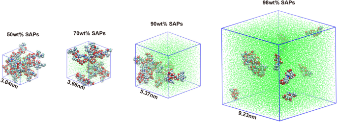
To discover the microstructure and water diffusion conduct, the polymer chain mannequin of SAPs was designed and the fractional free quantity (FFV) mannequin of the designed polymer chain was simulated the by molecular dynamics [28] (Fig. 2). The behaviour of the FFV with respect to the simulation time is illustrated in Determine S7A. The free quantity fraction will increase with growing water content material as a result of solubilising impact of SAPs on water uptake. The 98% saturated water content material mannequin for SAPs has the best free quantity fraction, averaging about 43%.
Using molecular dynamics simulation, the variety of hydrogen bonds fashioned between SAPs and water was quantified, in an effort to assess the hydration capability of SAPs (Determine S7B). It was discovered that the variety of hydrogen bonds between SAPs and water elevated with the growing water content material. In fashions containing 50wt% and 70wt% water content material, after 5000 ps, the variety of hydrogen bonds continued to rise over time. This improve was attributed to the predominantly secure nature of certain water inside SAPs. Nonetheless, at larger water contents of 90wt% or 98wt%, whereas the preliminary variety of hydrogen bonds remained excessive, a reducing pattern was noticed over the simulation interval. This lower was probably as a result of saturation of certain water websites and the emergence of free water molecules, which exhibit weaker binding interactions with SAPs. The interplay power of the SAPs with water was divided into Lennard–Jones potential (LJ, nonelectrostatic pressure) and Coulomb potential (electrostatic pressure) (Determine S7C and S7D). The interplay power for water of SAPs, was primarily supplied by the electrostatic gravitational pressure of the Coulomb potential. The change of Coulomb potential power of fashions with completely different water contents is in line with the hydrogen bond over the simulation time. In different phrases, the hydration means of SAPs was endowed by the cost impact that facilitates the attraction of water molecules to type extra hydrogen bonds with hydrophilic teams.
Cost neutralization of sodium polyacrylate (SP) and isolation of MSC-EVs by the IOSCE
EVs are negatively charged as a consequence of their phospholipid bilayer membranes, which permits them to be aggregated and precipitated by charge-based precipitation. On this research, SP was chosen to isolate MSC-EVs by cost neutralization. As proven in Determine S8, SP successfully removes water molecules surrounding the EVs, disrupting their hydration layer and growing their hydrophobicity, which in flip results in EV aggregation and precipitation [29, 30]. WB evaluation revealed that EV markers remoted utilizing 4% SP exhibited larger sign depth in comparison with these remoted with different SP focus (Determine S9). NTA evaluation confirmed that MSC-EVs remoted utilizing 4% SP had a mean dimension of 159.5 ± 64.7 nm, with a narrower distribution vary than these obtained with different concentrations, suggesting a extra uniform vesicle dimension distribution inside the samples (Figures S10, S11, Desk S2). Primarily based on these findings, 4% SP was chosen for subsequent experiments to isolate MSC-EVs together with SAPs focus. Moreover, the 4% SP proved efficient in separating EVs from plasma (Determine S12 and S13).
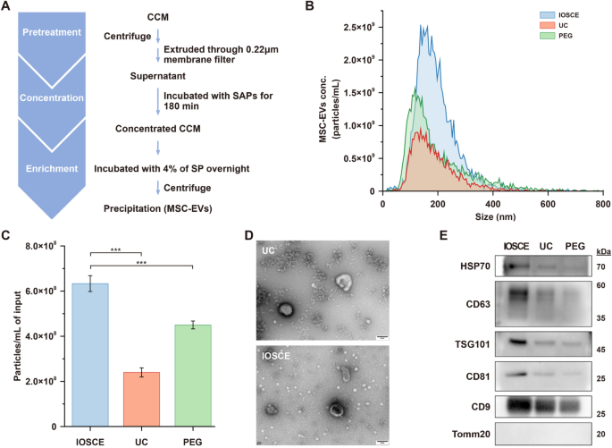
To evaluate our proposed IOSCE technique, CCM harvested from human umbilical twine MSCs was handled with the mix of SAP-0.75 and 4% SP (Fig. 3A). This was in comparison with UC and PEG-based precipitation strategies (20% PEG-6000) (Desk S3). The median dimension of the remoted particles of IOSCE was 176.8 ± 75.4 nm, exhibiting no important distinction in comparison with the opposite two strategies (UC = 164.7 ± 81.5 nm; 20% PEG = 154.3 ± 115 nm), all inside the dimension vary of small EVs (Fig. 3B). The yields of the MSC-EVs remoted by IOSCE was (6.33 ± 0.35) × 108 particles/mL, which is 2.64 occasions and 1.4 occasions larger than these remoted by UC and 20% PEG, respectively (Fig. 3C). As a result of decrease centrifugal pressure, MSC-EVs remoted by IOSCE exhibit a extra typical cup-shaped morphology in comparison with these obtained by UC (Fig. 3D). These findings point out that the IOSCE, successfully isolates MSC-EVs inside the attribute dimension vary, and achieves a better yield of MSC-EVs with extra intact morphology.
Isolation of MSC-EVs through the use of the IOSCE, UC and PEG. A Stream chart of MSC-EVs isolation by the IOSCE. B Particle dimension distributions and focus of MSC-EVs remoted by completely different strategies from CCM. The particle concentrations have been corrected for pattern enter volumes. C Particle yields of every technique. Particle yields have been corrected for pattern enter volumes. D TEM photographs of MSC-EVs obtained by UC and the IOSCE. Scale bars = 100 nm. E Western blots of HSP70, CD63, TSG101, CD81, CD9 and Tomm20 for MSC-EVs remoted from CCM by completely different strategies
Furthermore, the outcomes obtained from WB additionally proved the identical conclusion. EVs had been remoted from the identical quantity of CCM utilizing three completely different strategies, and the protein contents of CD9, CD81, CD63, TSG101, HSP70 and Tomm20 had been in contrast. As proven in Fig. 3E, the protein expression ranges of canonical EV markers (CD9, CD81, CD63, TSG101 and HSP70) had been considerably larger within the IOSCE strategies, indicating that IOSCE can yield extra EVs from the identical quantity of CCM. Moreover, the absence of the unfavorable marker (Tomm20) sign in EVs indicated the elimination of mobile impurities. To guage the purity of MSC-EVs enriched utilizing the IOSCE technique, we examined two key indicators: the particle-to-protein ratio and the RNA-to-protein ratio. As proven in Desk S4 and S5, the MSC-EVs enriched by IOSCE had a particle-to-protein ratio of (7.22 ± 0.4) × 1010 particles/μg and an RNA-to-protein ratio of 129.5 ± 10.2 pg/μg. In comparison with conventional strategies similar to ultracentrifugation (UC), dimension exclusion chromatography (SEC), and polymer precipitation, the IOSCE technique achieved superior purity ranges.
The IOSCE technique was employed to counterpoint MSC-EVs from 500 mL of CCM in an effort to assess the scalability of this strategy. As proven in Determine S14A and D, the particle dimension of EVs enriched from the massive pattern quantity was inside the anticipated nanometer vary, with a attribute cup-shaped morphology, in line with the requirements for small EVs. As depicted in Determine S14B, the MSC-EV yield was (6.43 ± 0.12) × 108/mL. Each particle dimension distribution and yield had been in line with the outcomes obtained from smaller pattern volumes. Furthermore, we characterised the everyday small EV markers, CD9, TSG101, and CD63, through WB, all of which produced clear bands, and there’s no contaminating protein Tomm20, a mitochondrial marker (Determine S14C).
These findings underscore the efficacy and scalability of IOSCE for the maximal focus of CCM and the environment friendly isolation of EVs, suggesting that IOSCE is a simpler technique than UC and precipitation strategies for isolating EVs from CCM.
Analysis of inhibiting impact of MSC-EVs remoted by the IOSCE on LPS-induced neuroinflammation in microglial cells
On this research, the anti-inflammatory impact of MSC-EVs remoted utilizing the IOSCE was investigated utilizing mouse microglial cell traces (BV2 cells). Previous to this, in vitro cytotoxicity and hemolysis assays had been performed on MSC-EVs obtained through the IOSCE technique to evaluate their security and biocompatibility. Enriched MSC-EVs had been first incubated with BV2 cells, with teams divided based mostly on various doses, and cell viability was monitored. As proven in Determine S15A, Calcein-AM/EthD-1 double staining revealed sturdy viability and progress in each the dosing and management teams, with minimal cell loss of life (purple fluorescence indicating EthD-1-staining useless cells). Moreover, CCK-8 assays had been employed to evaluate the results of MSC-EVs on BV2 cell progress. As proven within the Determine S15B, BV2 cells within the dosing teams exhibited strong viability in comparison with the NC group, with values of 124.34 ± 8.06%, 120.67 ± 10.12%, 118.02 ± 13.04%, and 97.06 ± 3.28%, respectively. Moreover, the hemolysis assay demonstrated no observable hemolytic exercise at MSC-EVs concentrations as much as 60 μg/mL (Determine S15C). Taken collectively, these outcomes point out that the enriched MSC-EVs exhibit wonderful biosafety, offering a strong basis for downstream purposes and the event of medical therapies.
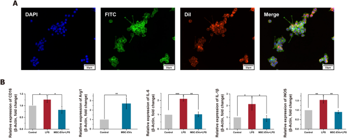
To determine a neuroinflammation mannequin, LPS-induced BV2 cells had been used, as LPS can stimulate the discharge of inflammatory cytokines in numerous cell sorts. The optimum therapeutic focus of MSC-EVs was first explored. As proven in Determine S16, cell viability progressively elevated because the focus of MSC-EVs rose from 0 μg/mL to 60 μg/mL. The utmost viability was noticed at a focus of 30 μg/mL, after which there was a slight lower. Due to this fact, we decided that 30 μg/mL of MSC-EVs because the optimized focus for subsequent experiments. The MSC-EVs had been successfully internalized by the BV2 cells (Fig. 4A). Microglia, as tissue-specific macrophages, share frequent macrophage traits and exist in two distinct activation states: the classical pro-inflammatory M1 state and the anti-inflammatory M2 state [31]. To elucidate the precise impact of LPS on microglial activation, the expression ranges of the M1 marker (CD16) and the M2 marker (Arg1) had been analyzed through qRT-PCR (Fig. 4B). The expressions of CD16 had been considerably upregulated after LPS remedy, however this upregulation was considerably diminished by pretreatment with MSC-EVs (p < 0.05). Conversely, Arg1 expression confirmed a major improve with MSC-EVs pretreatment (p < 0.01). These outcomes counsel that MSC-EVs can inhibit M1 microglial polarization whereas selling M2 polarization. Moreover, the degrees of pro-inflammatory elements, IL-6, IL-1β and iNOS, had been investigated utilizing qRT-PCR (Fig. 4B). MSC-EVs had been discovered to considerably scale back the discharge of those pro-inflammatory elements, a discovering additional corroborated by ELISA outcomes (Determine S17). This complete evaluation signifies the potential of MSC-EVs in modulating inflammatory responses in microglial cells.
The impact of MSC-EVs remoted by IOSCE on the lipopolysaccharides (LPS)-induced neuroinflammation. A Confocal photographs of the internalization of DiI-labeled MSC-EVs (purple) by BV2 cells. Nucleus and cytoskeleton of the BV2 cells had been labeled by 4’,6-diamidino-2-phenylindole (DAPI, blue) and fluorescein isothiocyanate (FITC, inexperienced), respectively. Scale bars = 50 μm. B mRNA ranges of CD16, Arg1, IL-6, IL-1β and iNOS as detected utilizing quantitative real-time PCR. Vital variations are indicated (*p < 0.05, **p < 0.01, ***p < 0.001)
Proteomic evaluation of the results of MSC-EVs on LPS-induced neuroinflammatory response in BV2 cells
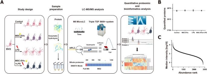
On this research, the results of MSC-EVs on neuroinflammation had been additional explored by conducting a high-throughput proteomic evaluation. Management, LPS-treated, MSC-EVs-treated and MSC-EVs + LPS-treated BV2 cells had been recognized through the use of DIA mass spectrometry. Knowledge unbiased acquisition (DIA) was chosen for its excessive information completeness and strong efficiency (Fig. 5A). A DIA library of about 3025 proteins was computationally merged from untreated and handled samples and a direct-DIA seek for all single-run samples. On common, 2863 proteins had been at a 1% peptide FDR quantified per pattern (Fig. 5B). The information acquired had excessive completeness, with 100% completeness for 2847 proteins (99.27%), 75% for 2867 proteins (99.93%), and 50% for 2867 proteins (99.97%) (Determine S18). A complete of 2847 proteins current in all runs had been used within the subsequent analyses. The relative abundance of the quantified proteins (normalized as iFOT) spanned six orders of magnitude after logarithmic transformation, which mirrored the deep sequencing protection (Fig. 5C, Determine S19).
Examine design and proteome characterization of the results of MSC-EVs on LPS-induced neuroinflammatory response in BV2 Cells. A Overview of the research teams and schematic proteomic workflow. B Variety of proteins recognized and quantified passing the 1% false discovery fee (FDR) cutoffs in every pattern. The dashed line signifies the imply of the quantified proteins. C Median protein abundance distribution of quantified proteins based mostly on iFOT intensities
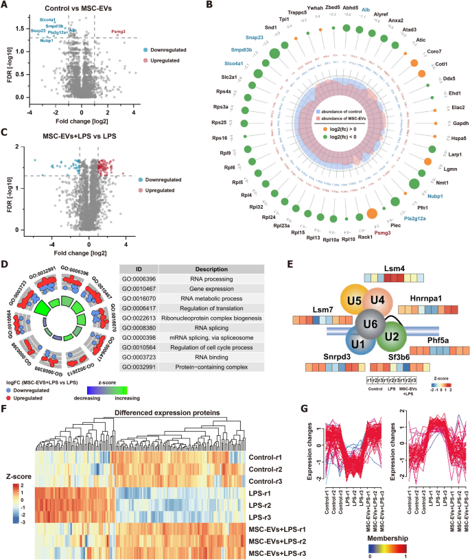
Subsequently, the impression of MSC-EVs on BV2 cells had been assessed by differential expression evaluation. Remarkably, exhibited no important change, with solely 7 proteins exhibiting important alterations [FDR < 0.05, Fold change (FC) > 2] (Fig. 6A). Notably, proteins within the NF-κB pathway, essential in mobile response to stimuli, confirmed no important adjustments. Nonetheless, the unfavorable regulator of this pathway, smpdl3b, was downregulated (Fig. 6B). The protein Psmg3, recognized to be downregulated in response to INF-γ and associated to inflammatory responses, was considerably upregulated in MSC-EVs internalized by BV2 cells, suggesting a possible anti-inflammatory position. This aligns with our phenotypic observations of a rise in M2-type BV2 cells, indicating that MSC-EVs minimally have an effect on protein expression in BV2 cells and should promote an anti-inflammatory state.
Differential proteomic evaluation and bioinformatics evaluation of the impression of MSC-EVs on BV2 cells. A Volcano plot for quantitative proteins between management and MSC-EVs-treated BV2 cells. The horizontal dashed line exhibits an FDR of 0.05. The vertical dashed traces present a fold change (FC) of two. B The differentially expressed proteins on management and MSC-EVs-treated BV2 cells (FDR < 0.05). The larger circle, the better the differential expression stage. Out to inside numbers in circles represented logarithmic values of the fold adjustments and the expression ranges of group management and MSC-EVs-treated BV2 cells. C Volcano plot for quantitative proteins between MSC-EVs + LPS-treated and LPS-treated BV2 cells. The horizontal dashed line exhibits an FDR of 0.05. The vertical dashed traces present an FC of two. D GO enrichment evaluation on differentially expressed proteins between MSC-EVs + LPS-treated and LPS-treated BV2 cells. E The protein expression ranges of six spliceosomal proteins of management, LPS-treated, and MSC-EVs + LPS-treated BV2 cells. F The heatmap evaluation of differential proteins between LPS vs. management and MSC-EVs + LPS vs. LPS. G The Mfuzz evaluation of differential proteins between LPS vs. management and MSC-EVs + LPS vs. LPS
Additional differential protein evaluation was performed to guage the results of LPS on BV2 cells and the anti-inflammatory potential of MSC-EVs. 105 proteins had been considerably modified after LPS stimulation (FDR < 0.05, FC > 2) (Determine S20), predominantly associated to RNA metabolic processes and regulation of translation (Determine S21). By evaluating the protein expression profiles of LPS-stimulated BV2 cells pretreated with and with out MSC-EVs, 113 proteins had been considerably modified (FDR < 0.05, FC > 2) (Fig. 6C). Apparently, the expression of functionalized differential proteins was additionally associated to RNA metabolic course of and regulation of translation, just like the organic processes concerned in differential proteins in BV2 cells earlier than and after LPS stimulation (Fig. 6D). Notably, MSC-EVs successfully countered important LPS-induced alterations in RNA metabolism and transcriptional regulatory proteins (Determine S22). For instance, MSC-EVs restored regular ranges of six spliceosomal proteins concerned in RNA splicing (Fig. 6E). A mixed evaluation of differential proteins between LPS vs. management and MSC-EVs + LPS vs. LPS by means of heatmap and Mfuzz analyses revealed that MSC-EVs broadly offset LPS-induced protein adjustments (Fig. 6F and G). Consequently, MSC-EVs seem to revive cells to their baseline state by reversing LPS-induced alterations in organic processes, emphasizing their potential in RNA metabolism and translational regulation modulation.


